Cysticercosis, MRI
Numéro d’image : 12636802
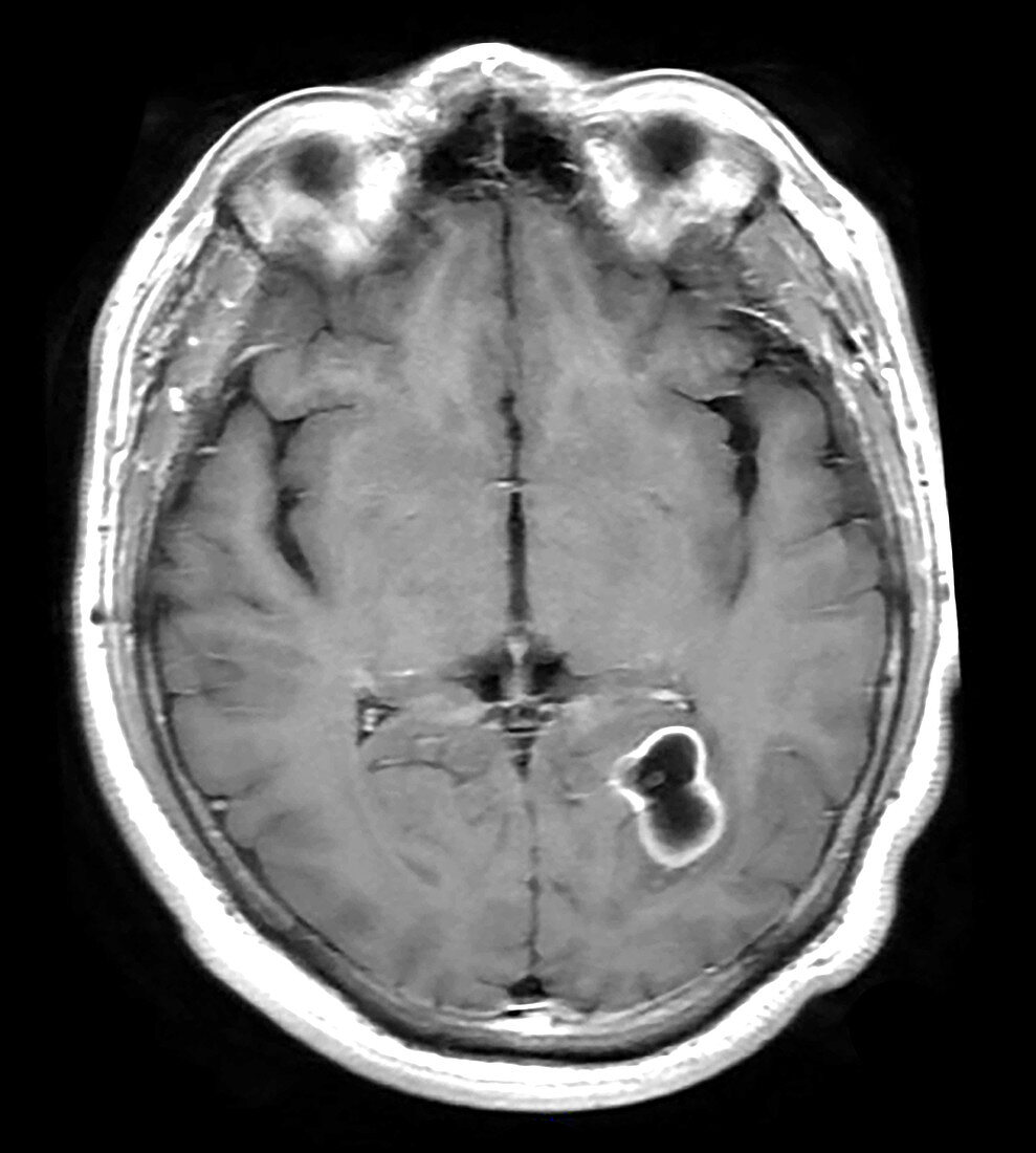
| This axial (cross section) T1 weighted MR image of the head with contrast enhancement shows a cystic lesion in the temporal-occipital region of the brain on the viewers right. The cyst is dark and the surrounding low signal is the inflammatory edema. There is peripheral enhancement along the wall of the cyst compatible with active infection. The small nodule within the cyst is the scolex (head of the pork tapeworm). This is a lesion related to infection from eggs of the pork tapeworm (taenia solium). This is the most common parasitic infection worldwide. The are parenchymal and extra-parenchymal forms of this infection. Multiple, different stages of infection can be determined best by MRI. | |
| Licence : | Droits gérés |
| Crédit: | Science Photo Library / Living Art Enterprises |
| Taille de l’image : | 3900 px × 4338 px |
| Model Release : | Non requis |
| Property Release : | Non requis |
| Restrictions : | - |
Prix pour cette image À partir de 45 €
Produit vendu
(Calendrier, Carte postale, Carte de vœux, Impression sur textile, Packaging etc)
À partir de 45 €
Usage commercial
(Affichage, Annonce presse, Annonce TV, Carte, Digital - hors rés. sociaux, Digital - rés. sociaux etc)
À partir de 45 €
Éditorial
(Digital, Journal, Livre, Livre pratique, Magazine, Télévision etc)
À partir de 60 €
Usage non-commercial
(Digital - hors rés. sociaux, Digital - rés. sociaux etc)
À partir de 120 €
Mots clés
- CYSTICERCOSIS,
- diagnostic,
- diagnostique,
- I.R.M.,
- imagerie médicale,
- imagerie par résonance magnétique,
- imagerie par résonnance magnétique,
- infection cérébrale,
- infection du cerveau,
- inflammation cérébrale,
- IRM,
- kyste au cerveau,
- kyste cérébral,
- médecine,
- médical,
- médicale,
- neurologie,
- neurologique,
- radiographie,
- système nerveux,
- TAENIA SOLIUM,
- ténia armé,
- ténia du porc