Cadaver Section through Brainstem
Numéro d’image : 12636734
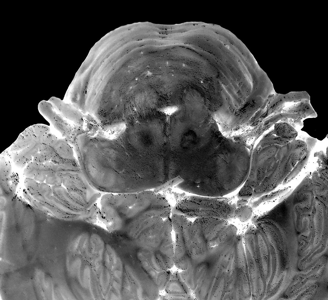
| This axial (cross sectional) slice of a cadaveric brain was performed at the level of the ponto-medullary junction. Black and white has been inverted. You are looking at the brainstem from below with the belly of the pons near the top of the image and the superior medulla below the pons. You can better visualize the internal structures within the brainstem. You can clearly see the proximal segments of the 7th and 8th cranial nerves. | |
| Licence : | Droits gérés |
| Crédit: | Science Photo Library / Living Art Enterprises |
| Taille de l’image : | 4259 px × 3900 px |
| Model Release : | Non requis |
| Property Release : | Non requis |
| Restrictions : | - |
Prix pour cette image À partir de 45 €
Produit vendu
(Calendrier, Carte postale, Carte de vœux, Impression sur textile, Packaging etc)
À partir de 45 €
Usage commercial
(Affichage, Annonce presse, Annonce TV, Carte, Digital - hors rés. sociaux, Digital - rés. sociaux etc)
À partir de 45 €
Éditorial
(Digital, Journal, Livre, Livre pratique, Magazine, Télévision etc)
À partir de 60 €
Usage non-commercial
(Digital - hors rés. sociaux, Digital - rés. sociaux etc)
À partir de 120 €