Prion Isoforms
Numéro d’image : 12636556
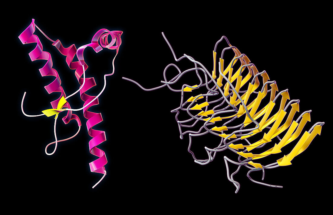
| A comparison of two 3D models of different prion protein isoforms. The structure on the left represents a human prion protein found on cell membranes in its normal state. The structure on the right is a fungal prion in it's folded amyloid form, similar to how prion proteins deform in certain neurological diseases like mad cow disease or Creutzfeldt-Jakob disease. | |
| Licence : | Droits gérés |
| Crédit: | Science Photo Library / Oto, Evan |
| Taille de l’image : | 5100 px × 3300 px |
| Model Release : | Non requis |
| Property Release : | Non requis |
| Restrictions : | - |
Prix pour cette image À partir de 45 €
Produit vendu
(Calendrier, Carte postale, Carte de vœux, Impression sur textile, Packaging etc)
À partir de 45 €
Usage commercial
(Affichage, Annonce presse, Annonce TV, Carte, Digital - hors rés. sociaux, Digital - rés. sociaux etc)
À partir de 45 €
Éditorial
(Digital, Journal, Livre, Livre pratique, Magazine, Télévision etc)
À partir de 60 €
Usage non-commercial
(Digital - hors rés. sociaux, Digital - rés. sociaux etc)
À partir de 120 €