Functional MRI, auditory stimulus
Numéro d’image : 12636548
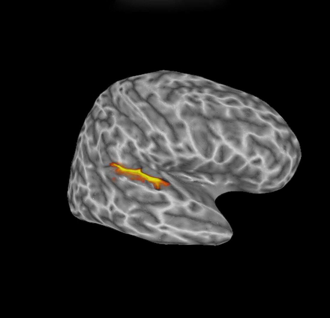
| Image shows the auditory cortex in the left hemisphere responding to a meaningful auditory stimulus (beyond the sounds of the MRI apparatus). The small, lighter coloured areas are selected based on p=0.05 and the wider areas (orange border) represent p = 0.1. Slight blur is due to the delay in the increased flow of blood to the stimulated areas. | |
| Licence : | Droits gérés |
| Crédit: | Science Photo Library / Bhatia, Kul |
| Taille de l’image : | 3287 px × 3162 px |
| Model Release : | Non requis |
| Property Release : | Non requis |
| Restrictions : | - |
Prix pour cette image À partir de 45 €
Produit vendu
(Calendrier, Carte postale, Carte de vœux, Impression sur textile, Packaging etc)
À partir de 45 €
Usage commercial
(Affichage, Annonce presse, Annonce TV, Carte, Digital - hors rés. sociaux, Digital - rés. sociaux etc)
À partir de 45 €
Éditorial
(Digital, Journal, Livre, Livre pratique, Magazine, Télévision etc)
À partir de 60 €
Usage non-commercial
(Digital - hors rés. sociaux, Digital - rés. sociaux etc)
À partir de 120 €