Submandibular Duct Stone, CT scan
Numéro d’image : 12628783
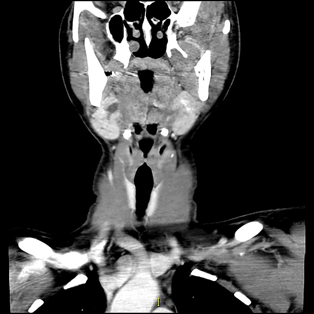
| This coronal (frontal) contrast enhanced CT image of the head and neck demonstrates dilatation of ducts within the submandibular gland secondary to an obstructing stone (calculus) in the floor of the mouth within the submandibular duct (Wharton's duct). | |
| Licence : | Droits gérés |
| Crédit: | Science Photo Library / Living Art Enterprises |
| Taille de l’image : | 3600 px × 3600 px |
| Model Release : | Non requis |
| Property Release : | Non requis |
| Restrictions : | - |
Prix pour cette image À partir de 45 €
Produit vendu
(Calendrier, Carte postale, Carte de vœux, Impression sur textile, Packaging etc)
À partir de 45 €
Usage commercial
(Affichage, Annonce presse, Annonce TV, Carte, Digital - hors rés. sociaux, Digital - rés. sociaux etc)
À partir de 45 €
Éditorial
(Digital, Journal, Livre, Livre pratique, Magazine, Télévision etc)
À partir de 60 €
Usage non-commercial
(Digital - hors rés. sociaux, Digital - rés. sociaux etc)
À partir de 120 €
Mots clés
- anormal,
- diagnostic,
- diagnostic par imagerie,
- diagnostique,
- diagnostique par imagerie,
- état,
- glande salivaire,
- glande sous maxillaire,
- glande sous-maxillaire,
- imagerie médicale,
- maladie,
- malsain,
- médical,
- médicale,
- pathologie,
- radiographie,
- radiologie,
- représentation,
- scanner,
- science,
- T.D.M.,
- TDM,
- tête et cou,
- tomodensitométrie