Hypothalamic Hamartoma, MRI
Numéro d’image : 12628768
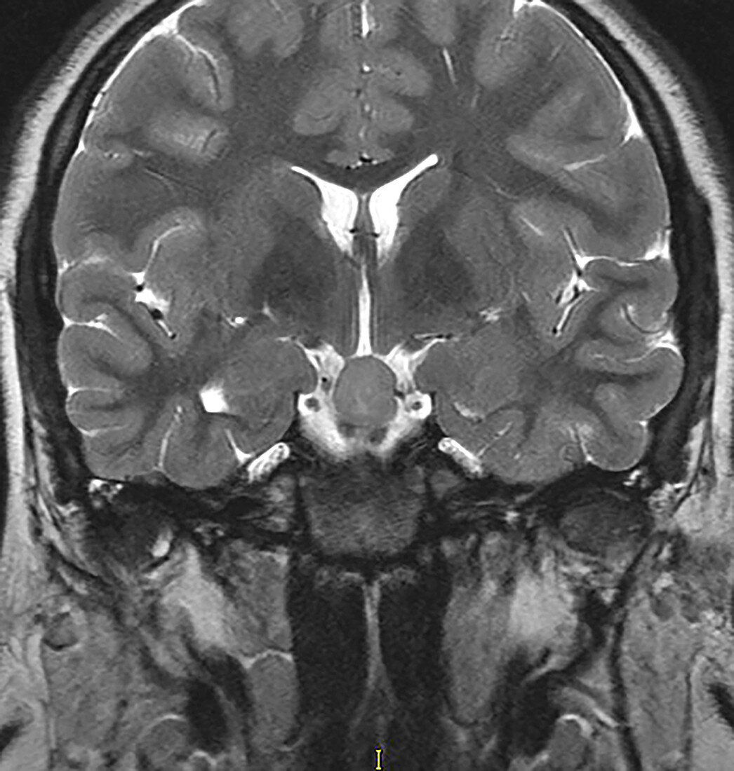
| This coronal (frontal) T2 weighted MR image of the head without contrast shows a well circumscribed isointense mass in the suprasellar cistern in this adolescent male with a history of gelastic (laughing) seizures. This is the typical appearance of a hypothalamic hamartoma. | |
| Licence : | Droits gérés |
| Crédit: | Science Photo Library / Living Art Enterprises |
| Taille de l’image : | 3600 px × 3785 px |
| Model Release : | Non requis |
| Property Release : | Non requis |
| Restrictions : | - |
Prix pour cette image À partir de 45 €
Produit vendu
(Calendrier, Carte postale, Carte de vœux, Impression sur textile, Packaging etc)
À partir de 45 €
Usage commercial
(Affichage, Annonce presse, Annonce TV, Carte, Digital - hors rés. sociaux, Digital - rés. sociaux etc)
À partir de 45 €
Éditorial
(Digital, Journal, Livre, Livre pratique, Magazine, Télévision etc)
À partir de 60 €
Usage non-commercial
(Digital - hors rés. sociaux, Digital - rés. sociaux etc)
À partir de 120 €
Mots clés
- anormal,
- cerveau,
- convulsions,
- crises,
- diagnostic,
- diagnostic par imagerie,
- diagnostique,
- diagnostique par imagerie,
- état,
- hamartome,
- I.R.M.,
- imagerie médicale,
- imagerie par résonance magnétique,
- imagerie par résonnance magnétique,
- IRM,
- IRM cérébrale,
- maladie,
- malsain,
- masse,
- médical,
- médicale,
- néoplasme,
- néoplasme cérébral,
- pathologie,
- radiographie,
- radiologie,
- représentation,
- réservoir,
- science,
- tumeur,
- tumeur cérébrale bénigne