Chronic Post-Traumatic Brain Injury, MRI
Numéro d’image : 12628461
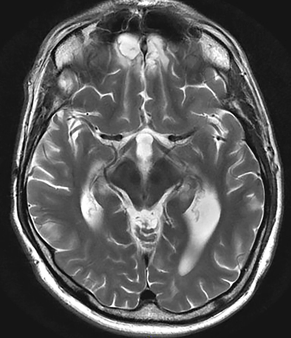
| This axial (cross sectional) T2 weighted MRI image demonstrates areas of chronic post-traumatic encephalomalacia (brain damage) in a typical location in the frontal lobes. Because the frontal lobes are so close to the adjacent bony skull base the brain hits the bone during the injury and then over time becomes atrophic and gliotic (brains response to injury). In the chronic phase the area of injury on MRI shows lower (looks darker) signal on T1 and higher (brighter or whiter) signal on T2 weighted images, as in this case. These brighter/whiter regions, partially represent cystic change of the brain due to the brain damage. | |
| Licence : | Droits gérés |
| Crédit: | Science Photo Library / Living Art Enterprises |
| Taille de l’image : | 3600 px × 4175 px |
| Model Release : | Non requis |
| Property Release : | Non requis |
| Restrictions : | - |
Prix pour cette image À partir de 45 €
Produit vendu
(Calendrier, Carte postale, Carte de vœux, Impression sur textile, Packaging etc)
À partir de 45 €
Usage commercial
(Affichage, Annonce presse, Annonce TV, Carte, Digital - hors rés. sociaux, Digital - rés. sociaux etc)
À partir de 45 €
Éditorial
(Digital, Journal, Livre, Livre pratique, Magazine, Télévision etc)
À partir de 60 €
Usage non-commercial
(Digital - hors rés. sociaux, Digital - rés. sociaux etc)
À partir de 120 €
Mots clés
- analyse transversale,
- athrophique,
- atrophie,
- axial,
- blessure,
- cerveau,
- chronique,
- diagnostic,
- diagnostique,
- dommage,
- gliotique,
- humain,
- I.R.M.,
- imagerie par résonance magnétique,
- imagerie par résonnance magnétique,
- IRM,
- médical,
- médicale,
- pondéré T2,
- post traumatic,
- post-traumatique,
- posttraumatique,
- scanner,
- transversale