Brain Anatomy, Illustration
Numéro d’image : 12628146
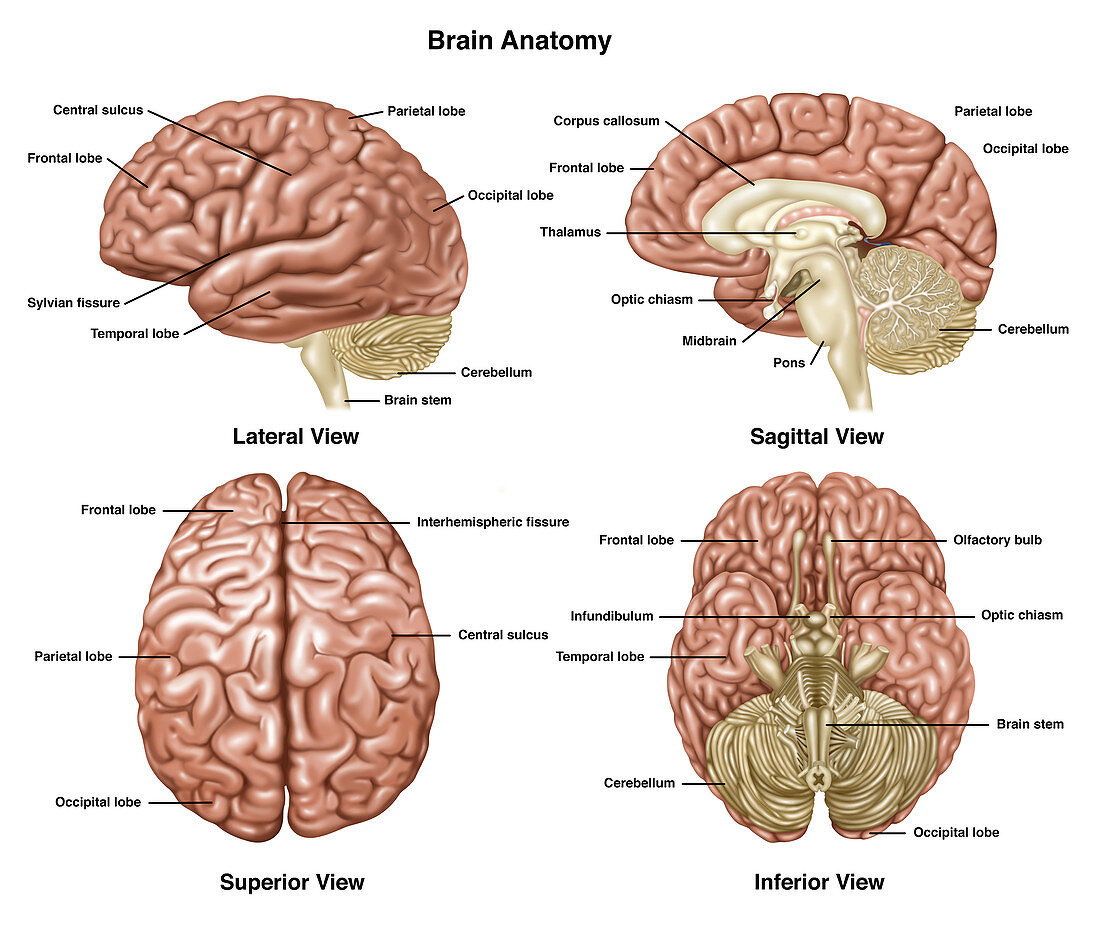
| Illustration showing anatomy of a normal brain in lateral, sagittal, superior, and inferior view. Notated are frontal lobe, central sulcus, parietal lobe, sylvian fissure, temporal lobe, occipital lobe, cerebellum, brain stem, corpus callosum, thalamus, optic chiasma, midbrain, pons, interhemispheric fissure, olfactory bulb, and infundibulum | |
| Licence : | Droits gérés |
| Crédit: | Science Photo Library / Shockey, Gwen |
| Taille de l’image : | 6300 px × 5400 px |
| Model Release : | Non requis |
| Property Release : | Non requis |
| Restrictions : | - |
Prix pour cette image À partir de 45 €
Produit vendu
(Calendrier, Carte postale, Carte de vœux, Impression sur textile, Packaging etc)
À partir de 45 €
Usage commercial
(Affichage, Annonce presse, Annonce TV, Carte, Digital - hors rés. sociaux, Digital - rés. sociaux etc)
À partir de 45 €
Éditorial
(Digital, Journal, Livre, Livre pratique, Magazine, Télévision etc)
À partir de 60 €
Usage non-commercial
(Digital - hors rés. sociaux, Digital - rés. sociaux etc)
À partir de 120 €
Mots clés
- ampoule olfactive,
- anatomie du cerveau,
- annoté,
- cerebellum,
- cerebrum,
- cerveau,
- cervelet,
- chiasme optique,
- corps calleux,
- corpus callosum,
- en bonne santé,
- graphique d'information,
- graphique informatif,
- illustration,
- infographie,
- infundibulum,
- lobe frontal,
- lobe occipital,
- lobe pariétal,
- lobe temporal,
- médical,
- médicale,
- mésencéphale,
- normal,
- oeuvre,
- plan sagittal,
- Pons,
- sain,
- scissure de Rolando,
- scissure de Sylvius,
- sillon central,
- sillon latéral,
- sulcature centrale,
- sulcus lateralis,
- thalamus,
- tronc cérébral,
- vue inférieure,
- vue latérale,
- vue sagittale,
- vue supérieure