Monomeric Ribosomes, TEM
Numéro d’image : 12627260
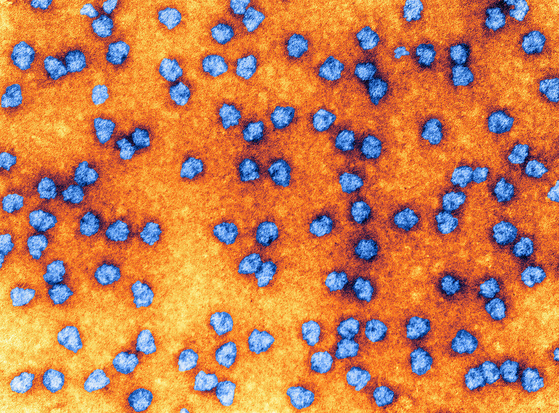
| Colour enhanced transmission electron micrograph of a field of monomeric ribosomes. Shown here are several frontal and lateral images. Most lateral images are of the right type and most frontal images are left-featured, but one is right-featured. An image which is intermediate between a right-featured frontal and left lateral image is also seen. Small subunit partitions are indicated by short arrows. Note that in left-featured frontal images the dense line in between subunits is not prominent towards the right side of the dense spot which is under the small subunit partition. Magnification 225, 000x 5x7 inches. | |
| Licence : | Droits gérés |
| Crédit: | Science Photo Library / Science Source |
| Taille de l’image : | 4210 px × 3107 px |
| Model Release : | Non requis |
| Property Release : | Non requis |
| Restrictions : | - |
Prix pour cette image À partir de 45 €
Produit vendu
(Calendrier, Carte postale, Carte de vœux, Impression sur textile, Packaging etc)
À partir de 45 €
Usage commercial
(Affichage, Annonce presse, Annonce TV, Carte, Digital - hors rés. sociaux, Digital - rés. sociaux etc)
À partir de 45 €
Éditorial
(Digital, Journal, Livre, Livre pratique, Magazine, Télévision etc)
À partir de 60 €
Usage non-commercial
(Digital - hors rés. sociaux, Digital - rés. sociaux etc)
À partir de 120 €
Mots clés
- amélioré,
- augmenté,
- biologie,
- biologie cellulaire,
- colorié,
- colorisé,
- histologie,
- M.E.,
- M.E.T.,
- machine moléculaire,
- ME,
- MET,
- micrographie,
- micrographie électronique,
- microscope,
- microscope électronique,
- microscope électronique à transmission,
- microscope électronique en transmission,
- microscopie,
- organite,
- renforcé,
- ribosome