Asian knotweed epidermis, light micrograph
Numéro d’image : 12584382
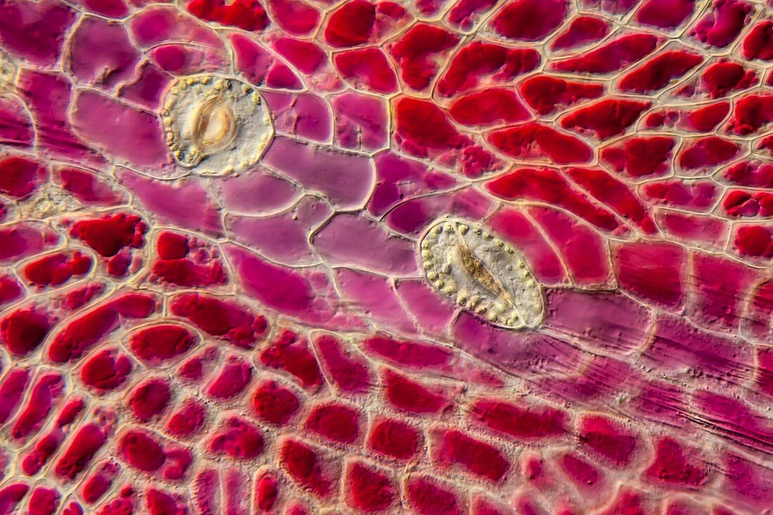
| Light micrograph of Asian knotweed epidermis. The image shows the stem epidermis of Asian knotweed, Fallopia japonica, taken from the red-painted areas of the stem epidermis. The colour is caused by the several cell vacuoles filled with anthocyanin. Two stomata are embedded in the epidermis cells. Microscopic contrast technique: Differential interference contrast. Magnification: 400x when printed 10 centimetres wide. | |
| Licence : | Droits gérés |
| Crédit: | Science Photo Library / Guenther, Gerd |
| Taille de l’image : | 5616 px × 3744 px |
| Model Release : | Non requis |
| Property Release : | Non requis |
| Restrictions : | - |
Prix pour cette image À partir de 45 €
Produit vendu
(Calendrier, Carte postale, Carte de vœux, Impression sur textile, Packaging etc)
À partir de 45 €
Usage commercial
(Affichage, Annonce presse, Annonce TV, Carte, Digital - hors rés. sociaux, Digital - rés. sociaux etc)
À partir de 45 €
Éditorial
(Digital, Journal, Livre, Livre pratique, Magazine, Télévision etc)
À partir de 60 €
Usage non-commercial
(Digital - hors rés. sociaux, Digital - rés. sociaux etc)
À partir de 120 €
Mots clés
- Asie,
- biologie,
- biologique,
- botanique,
- contraste d'interférence différentielle,
- contraste interférentiel différentiel,
- contratse différentiel d'interférence,
- épiderme,
- fallopia japonica,
- flore,
- microscope,
- microscope optique,
- microscopie,
- microscopie optique,
- microscopique,
- nature,
- stomates,
- vacuole