Liver haemochromatosis, light micrograph
Numéro d’image : 12583834
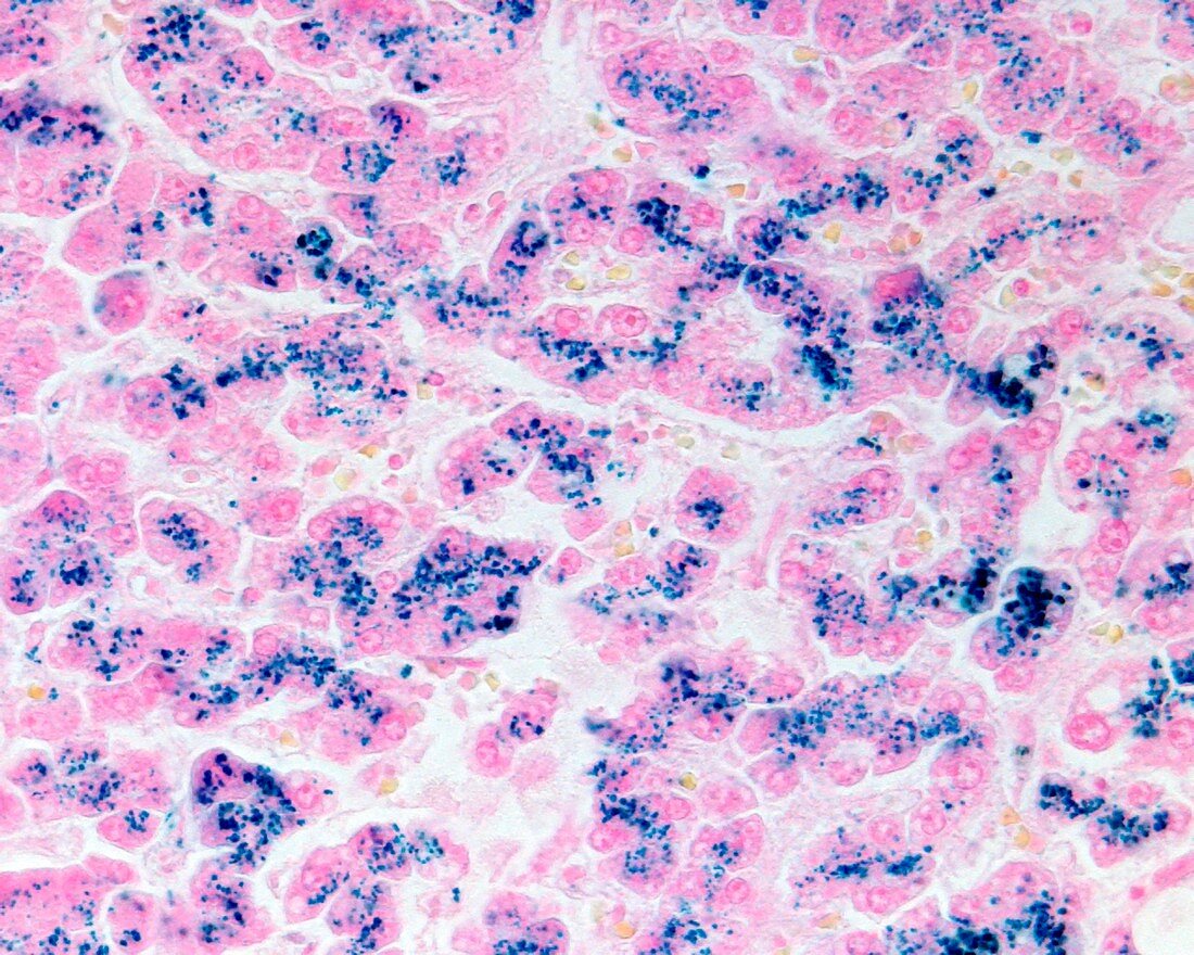
| Light micrograph of a liver biopsy stained with Perls' Prussian blue method showing iron deposits (blue). In haemochromatosis, excessive iron is deposited in hepatocytes and characteristically concentrated in the cytoplasm around bile canaliculi (parenchymal haemosiderosis). | |
| Licence : | Droits gérés |
| Crédit: | Science Photo Library / Jose Calvo |
| Taille de l’image : | 4674 px × 3739 px |
| Model Release : | Non requis |
| Property Release : | Non requis |
| Restrictions : | - |
Prix pour cette image À partir de 45 €
Produit vendu
(Calendrier, Carte postale, Carte de vœux, Impression sur textile, Packaging etc)
À partir de 45 €
Usage commercial
(Affichage, Annonce presse, Annonce TV, Carte, Digital - hors rés. sociaux, Digital - rés. sociaux etc)
À partir de 45 €
Éditorial
(Digital, Journal, Livre, Livre pratique, Magazine, Télévision etc)
À partir de 60 €
Usage non-commercial
(Digital - hors rés. sociaux, Digital - rés. sociaux etc)
À partir de 120 €
Mots clés
- biopsie,
- catégorie,
- cellule,
- cellules,
- corps humain,
- coupe,
- désordre,
- divisé,
- état,
- excédent,
- excès,
- foie,
- génétique,
- hématochromatose,
- hémochromatose,
- hépatique,
- héréditaire,
- histologie,
- histologique,
- malade,
- maladie,
- médecine,
- médical,
- médicale,
- microscope,
- microscope optique,
- microscopie optique,
- partie,
- pathologie,
- pathologique,
- récessif,
- section,
- tissus,
- trouble