Liver haemochromatosis, light micrograph
Numéro d’image : 12583833
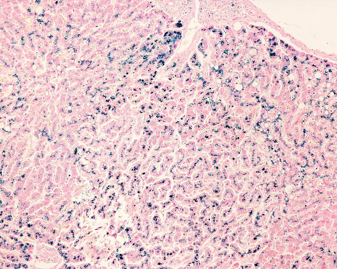
| Light micrograph of a liver biopsy stained with Perls' Prussian blue method showing iron deposits (blue). In hereditary haemochromatosis the iron deposits are not homogeneous, leaving some iron-free areas (right and left border of image). In haemochromatosis, excessive iron is deposited in hepatocytes and characteristically concentrated in the cytoplasm around bile canaliculi. | |
| Licence : | Droits gérés |
| Crédit: | Science Photo Library / Jose Calvo |
| Taille de l’image : | 4674 px × 3739 px |
| Model Release : | Non requis |
| Property Release : | Non requis |
| Restrictions : | - |
Prix pour cette image À partir de 45 €
Produit vendu
(Calendrier, Carte postale, Carte de vœux, Impression sur textile, Packaging etc)
À partir de 45 €
Usage commercial
(Affichage, Annonce presse, Annonce TV, Carte, Digital - hors rés. sociaux, Digital - rés. sociaux etc)
À partir de 45 €
Éditorial
(Digital, Journal, Livre, Livre pratique, Magazine, Télévision etc)
À partir de 60 €
Usage non-commercial
(Digital - hors rés. sociaux, Digital - rés. sociaux etc)
À partir de 120 €
Mots clés
- biopsie,
- catégorie,
- cellule,
- cellules,
- corps humain,
- coupe,
- désordre,
- divisé,
- état,
- excédent,
- excès,
- foie,
- génétique,
- hématochromatose,
- hémochromatose,
- hépatique,
- héréditaire,
- histologie,
- histologique,
- malade,
- maladie,
- médecine,
- médical,
- médicale,
- microscope,
- microscope optique,
- microscopie optique,
- partie,
- pathologie,
- pathologique,
- récessif,
- section,
- tissus,
- trouble