Didinium attacking Paramecium
Numéro d’image : 12528439
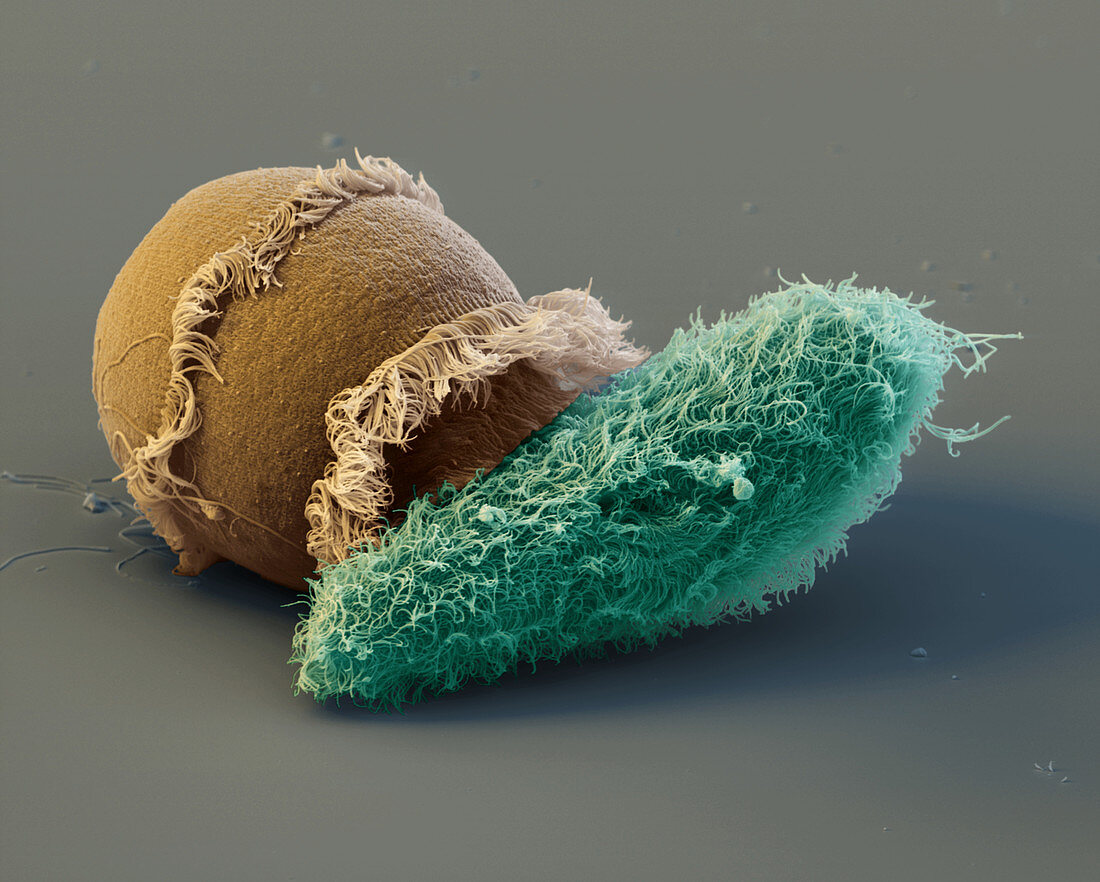
| Didinium attacks Paramecium. Coloured scanning electron micrograph (SEM) showing a one-celled Didinium (brown) in the process of attacking a one-celled Paramecium sp. (blue). These animals are ciliate protozoans, both bearing cilia. The barrel-shaped Didinium has two girdles of loco- motory cilia, and a prominent snout which is used as a probing and seizing organ during feeding. The Paramecium has cilia across its body used for movement. The Didinium has manoeuvred the Para- mecium, almost twice its own size, into an easier position for ingestion. The Didinium's snout and gullet are expanding in preparation for the meal. Magnification: x600 at 6x7cm size. | |
| Licence : | Droits gérés |
| Crédit: | Science Photo Library / EYE OF SCIENCE |
| Taille de l’image : | 2000 px × 1603 px |
| Model Release : | Non requis |
| Property Release : | Non requis |
| Restrictions : |
|
Prix pour cette image À partir de 45 €
Produit vendu
(Calendrier, Carte postale, Carte de vœux, Impression sur textile, Packaging etc)
À partir de 45 €
Usage commercial
(Affichage, Annonce presse, Annonce TV, Carte, Digital - hors rés. sociaux, Digital - rés. sociaux etc)
À partir de 45 €
Éditorial
(Digital, Journal, Livre, Livre pratique, Magazine, Télévision etc)
À partir de 60 €
Usage non-commercial
(Digital - hors rés. sociaux, Digital - rés. sociaux etc)
À partir de 120 €