Muscle fibres, SEM
Numéro d’image : 12528147
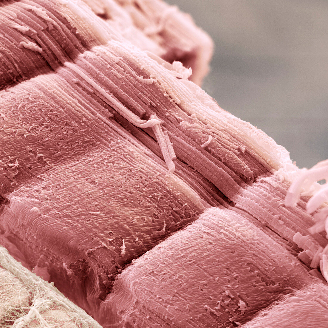
| Skeletal muscle. Coloured scanning electron micrograph (SEM) of skeletal (striated) muscle fibres. The muscle fibres are extremely long, cylindrical cells. Running the length of each fibre are thin threads called myofibrils (top left to bottom right), which contain the proteins actin and myosin. These proteins slide over each other to allow the muscle to contract. The alternating bands of actin and myosin give the fibres a striped appearance. Skeletal muscle is attached to the skeleton and is under the control of the voluntary nervous system. Magnification: x2000 when printed 10 centimetres wide. | |
| Licence : | Droits gérés |
| Crédit: | Science Photo Library / EYE OF SCIENCE |
| Taille de l’image : | 2000 px × 2000 px |
| Model Release : | Non requis |
| Property Release : | Non requis |
| Restrictions : |
|
Prix pour cette image À partir de 45 €
Produit vendu
(Calendrier, Carte postale, Carte de vœux, Impression sur textile, Packaging etc)
À partir de 45 €
Usage commercial
(Affichage, Annonce presse, Annonce TV, Carte, Digital - hors rés. sociaux, Digital - rés. sociaux etc)
À partir de 45 €
Éditorial
(Digital, Journal, Livre, Livre pratique, Magazine, Télévision etc)
À partir de 60 €
Usage non-commercial
(Digital - hors rés. sociaux, Digital - rés. sociaux etc)
À partir de 120 €