Posterior pituitary gland. Herring body, TEM
Numéro d’image : 12504731
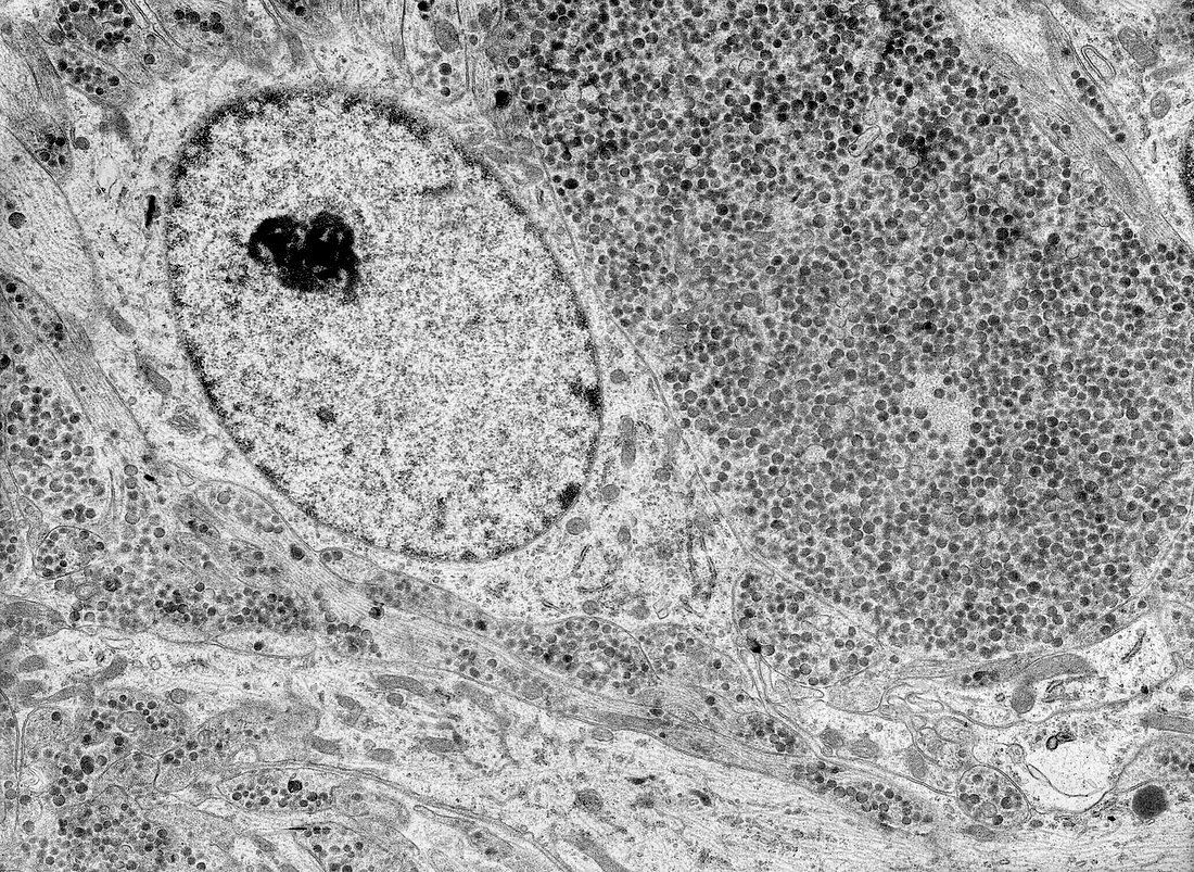
| Transmission electron micrograph (TEM) of the pituitary posterior lobe showing an axonal swelling loaded with neurosecretory granules known as Herring bodies (dark dots) and a pituicyte glial cell (centre left). | |
| Licence : | Droits gérés |
| Crédit: | Science Photo Library / JOSE CALVO |
| Taille de l’image : | 3790 px × 2766 px |
| Model Release : | Non requis |
| Property Release : | Non requis |
| Restrictions : | - |
Prix pour cette image À partir de 45 €
Produit vendu
(Calendrier, Carte postale, Carte de vœux, Impression sur textile, Packaging etc)
À partir de 45 €
Usage commercial
(Affichage, Annonce presse, Annonce TV, Carte, Digital - hors rés. sociaux, Digital - rés. sociaux etc)
À partir de 45 €
Éditorial
(Digital, Journal, Livre, Livre pratique, Magazine, Télévision etc)
À partir de 60 €
Usage non-commercial
(Digital - hors rés. sociaux, Digital - rés. sociaux etc)
À partir de 120 €
Mots clés
- aucun,
- cellule,
- cytologie,
- cytologique,
- glande endocrinale,
- glande endocrine,
- histologie,
- histologique,
- hypophyse,
- hypophyse postérieure,
- lobe arrière,
- lobe postérieur,
- M.E.T.,
- MET,
- micrographie électronique à transmission,
- microscope,
- microscope électronique à transmission,
- microscopie,
- neurohypophyse,
- personne,
- posthypophyse,
- ultrastructure