Glial limiting membrane, TEM
Numéro d’image : 12504725
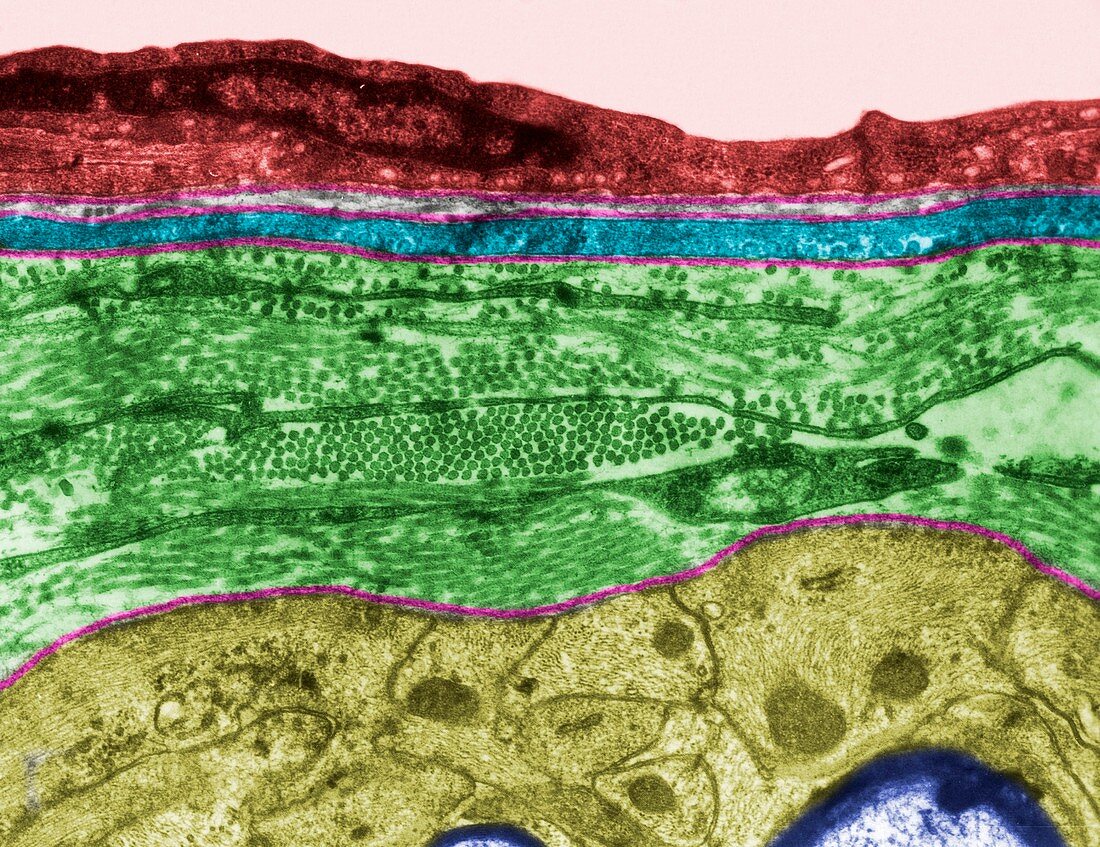
| Coloured transmission electron micrograph (TEM) showing the glial limiting membrane (glia limitans) of white matter. It is formed by astrocyte processes (yellow) that separated blood capillary endothelium (red), pericyte (blue) and the surrounding connective tissue (green) from the myelinated nerve fibres (dark purple, bottom). Basal lamina is pink. | |
| Licence : | Droits gérés |
| Crédit: | Science Photo Library / JOSE CALVO |
| Taille de l’image : | 3622 px × 2790 px |
| Model Release : | Non requis |
| Property Release : | Non requis |
| Restrictions : | - |
Prix pour cette image À partir de 45 €
Produit vendu
(Calendrier, Carte postale, Carte de vœux, Impression sur textile, Packaging etc)
À partir de 45 €
Usage commercial
(Affichage, Annonce presse, Annonce TV, Carte, Digital - hors rés. sociaux, Digital - rés. sociaux etc)
À partir de 45 €
Éditorial
(Digital, Journal, Livre, Livre pratique, Magazine, Télévision etc)
À partir de 60 €
Usage non-commercial
(Digital - hors rés. sociaux, Digital - rés. sociaux etc)
À partir de 120 €
Mots clés
- astrocyte,
- aucun,
- basal lamina,
- biologie,
- biologique,
- capillaire,
- cellule,
- coloré,
- colorié,
- colorisé,
- cytologie,
- cytologique,
- glie,
- histologie,
- histologique,
- M.E.T.,
- MET,
- micrographie électronique à transmission,
- microscope,
- microscope électronique à transmission,
- microscopie,
- personne,
- S.N.C.,
- SNC,
- système nerveux central,
- tissu conjonctif,
- ultrastructure,
- ultrastructure des cellules