Langhans cell in tuberculosis, light micrograph
Numéro d’image : 12491675
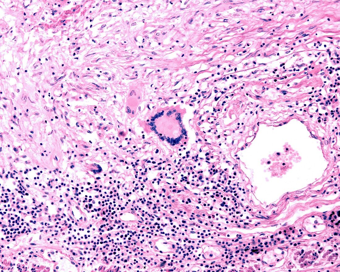
| Light micrograph of a Langhans-type giant cell in the periphery of a tuberculosis granuloma (inflamed nodule). In the lower half is a chronic inflammatory infiltrate. In the upper half is an area of fibrosis. Magnification: x180 when printed at 10 centimetres wide. | |
| Licence : | Droits gérés |
| Crédit: | Science Photo Library / JOSE CALVO |
| Taille de l’image : | 4674 px × 3739 px |
| Model Release : | Non requis |
| Property Release : | Non requis |
| Restrictions : | - |
Prix pour cette image À partir de 45 €
Produit vendu
(Calendrier, Carte postale, Carte de vœux, Impression sur textile, Packaging etc)
À partir de 45 €
Usage commercial
(Affichage, Annonce presse, Annonce TV, Carte, Digital - hors rés. sociaux, Digital - rés. sociaux etc)
À partir de 45 €
Éditorial
(Digital, Journal, Livre, Livre pratique, Magazine, Télévision etc)
À partir de 60 €
Usage non-commercial
(Digital - hors rés. sociaux, Digital - rés. sociaux etc)
À partir de 120 €
Mots clés
- anormal,
- corps humain,
- désordre,
- état,
- histopathologie,
- histopathologique,
- infiltrez,
- inflammatoire,
- maladie,
- malsain,
- médecine,
- médical,
- médicale,
- micrographie,
- micrographie optique,
- microscope,
- microscope optique,
- microscopie,
- microscopie optique,
- pathologie,
- poumon,
- pulmonaire,
- système respiratoire,
- trouble