Layers of trachea wall, light micrograph
Numéro d’image : 12490957
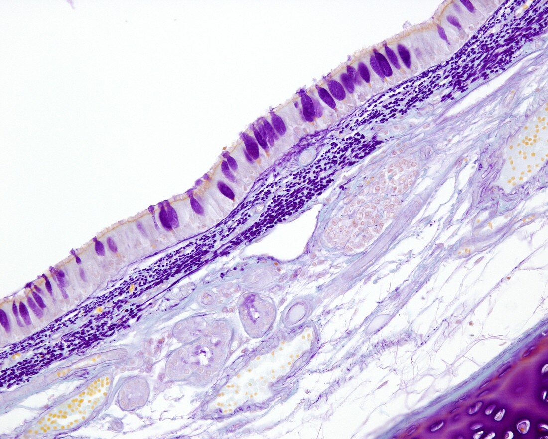
| Layers of trachea wall, light micrograph. At left is the mucosa layer, which consists of respiratory epithelium (uppermost) with goblet cells (purple ovals) and the lamina propria with elastic fibres (dark purple dots) and blood vessels (filled with yellow red blood cells). At right is the hyaline cartilage ring (red) and then the adventitia layer. Gabe technique. Magnification: x90 when printed at 10 centimetres wide. | |
| Licence : | Droits gérés |
| Crédit: | Science Photo Library / JOSE CALVO |
| Taille de l’image : | 4674 px × 3739 px |
| Model Release : | Non requis |
| Property Release : | Non requis |
| Restrictions : | - |
Prix pour cette image À partir de 45 €
Produit vendu
(Calendrier, Carte postale, Carte de vœux, Impression sur textile, Packaging etc)
À partir de 45 €
Usage commercial
(Affichage, Annonce presse, Annonce TV, Carte, Digital - hors rés. sociaux, Digital - rés. sociaux etc)
À partir de 45 €
Éditorial
(Digital, Journal, Livre, Livre pratique, Magazine, Télévision etc)
À partir de 60 €
Usage non-commercial
(Digital - hors rés. sociaux, Digital - rés. sociaux etc)
À partir de 120 €