Trachea and oesophagus, light micrograph
Numéro d’image : 12490948
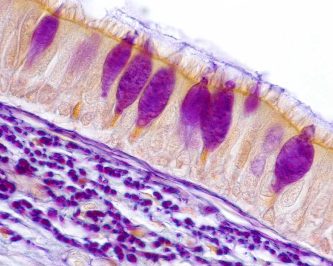
| Very low magnification light micrograph showing the neck organs in cross-section. The front of the neck is at right. The large space at centre is the trachea (windpipe) To its right is the thyroid cartilage (dark purple) and thyroid gland. The squashed space at left is the oesophagus. Surrounding the organs are muscles and adipose tissue. Magnification: x1 when printed at 10 centimetres wide. | |
| Licence : | Droits gérés |
| Crédit: | Science Photo Library / JOSE CALVO |
| Taille de l’image : | 4674 px × 3739 px |
| Model Release : | Non requis |
| Property Release : | Non requis |
| Restrictions : | - |
Prix pour cette image À partir de 45 €
Produit vendu
(Calendrier, Carte postale, Carte de vœux, Impression sur textile, Packaging etc)
À partir de 45 €
Usage commercial
(Affichage, Annonce presse, Annonce TV, Carte, Digital - hors rés. sociaux, Digital - rés. sociaux etc)
À partir de 45 €
Éditorial
(Digital, Journal, Livre, Livre pratique, Magazine, Télévision etc)
À partir de 60 €
Usage non-commercial
(Digital - hors rés. sociaux, Digital - rés. sociaux etc)
À partir de 120 €
Mots clés
- adipeux,
- anneaux,
- aucun,
- biologie,
- biologique,
- cartilage,
- cou,
- en bonne santé,
- éosine,
- hématoxyline,
- histologie,
- histologique,
- hyalin,
- micrographie,
- micrographie optique,
- microscope,
- microscope optique,
- microscopie,
- microscopie optique,
- mucosa,
- muqueuse,
- normal,
- œsophage,
- oesophagus,
- personne,
- presque transparent,
- respiratoire,
- sain,
- thyroïde,
- trachea,
- trachée,
- translucide