Layers of trachea wall, light micrograph
Numéro d’image : 12490946
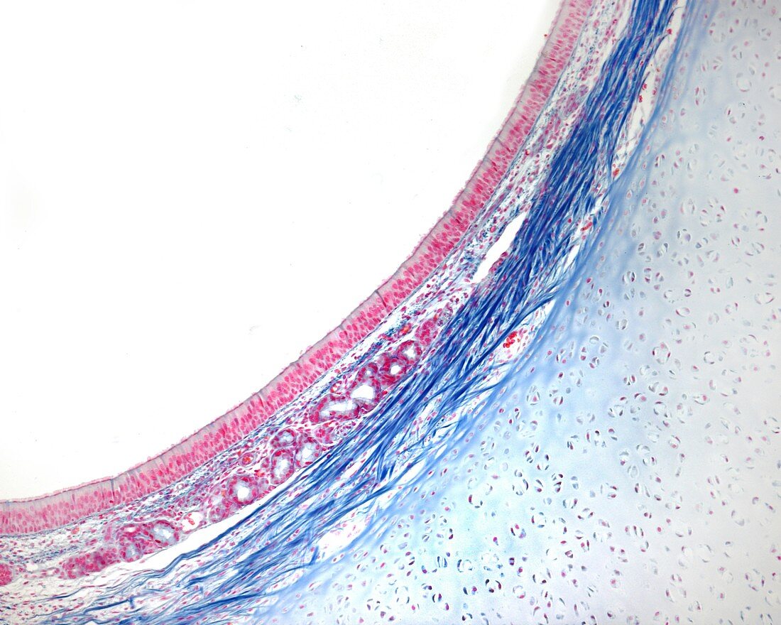
| Layers of trachea wall, light micrograph. The uppermost layer is the mucosa, which is lined by pseudostratified columnar epithelium (respiratory epithelium). Below this is the lamina propria, which contains blood vessels and mucus glands. At right is hyaline cartilage. Collagen fibres appear deep blue. Pasini method. Magnification: x90 when printed at 10 centimetres wide. | |
| Licence : | Droits gérés |
| Crédit: | Science Photo Library / JOSE CALVO |
| Taille de l’image : | 4674 px × 3739 px |
| Model Release : | Non requis |
| Property Release : | Non requis |
| Restrictions : | - |
Prix pour cette image À partir de 45 €
Produit vendu
(Calendrier, Carte postale, Carte de vœux, Impression sur textile, Packaging etc)
À partir de 45 €
Usage commercial
(Affichage, Annonce presse, Annonce TV, Carte, Digital - hors rés. sociaux, Digital - rés. sociaux etc)
À partir de 45 €
Éditorial
(Digital, Journal, Livre, Livre pratique, Magazine, Télévision etc)
À partir de 60 €
Usage non-commercial
(Digital - hors rés. sociaux, Digital - rés. sociaux etc)
À partir de 120 €