Skeletal muscle fibres, light micrograph
Numéro d’image : 12490216
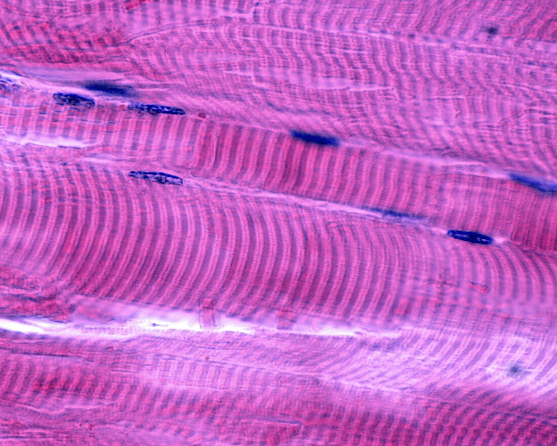
| Skeletal muscle fibres in cross section, light micrograph. The dotted appearance is due to myofibrils composed of bundles of myosin and actin filaments. The nuclei are displaced to the periphery of the muscle cell. Magnification: x900 when printed at 10 centimetres wide. | |
| Licence : | Droits gérés |
| Crédit: | Science Photo Library / JOSE CALVO |
| Taille de l’image : | 3840 px × 3072 px |
| Model Release : | Non requis |
| Property Release : | Non requis |
| Restrictions : | - |
Prix pour cette image À partir de 45 €
Produit vendu
(Calendrier, Carte postale, Carte de vœux, Impression sur textile, Packaging etc)
À partir de 45 €
Usage commercial
(Affichage, Annonce presse, Annonce TV, Carte, Digital - hors rés. sociaux, Digital - rés. sociaux etc)
À partir de 45 €
Éditorial
(Digital, Journal, Livre, Livre pratique, Magazine, Télévision etc)
À partir de 60 €
Usage non-commercial
(Digital - hors rés. sociaux, Digital - rés. sociaux etc)
À partir de 120 €
Mots clés
- aucun,
- bande I,
- biologie,
- biologique,
- cellule,
- en bonne santé,
- fibre,
- histologie,
- histologique,
- I,
- langue,
- ligne Z,
- micrographie,
- micrographie optique,
- microscope,
- microscope optique,
- microscopie,
- microscopie optique,
- muscle,
- musculaire,
- myocyte,
- myofibrille,
- normal,
- personne,
- sain,
- sarcomère,
- squelettique,
- strié,
- tissus,
- une bande