Lysosomes in a kidney, light micrograph
Numéro d’image : 12448076
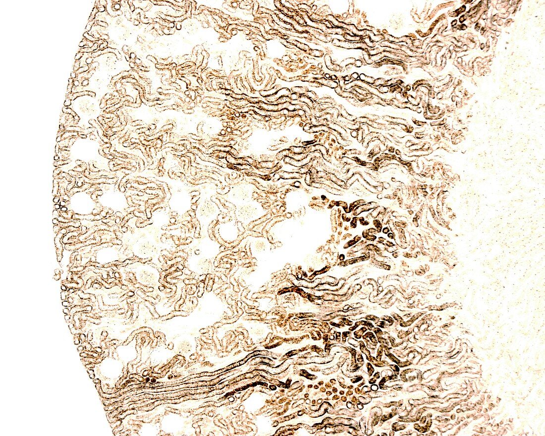
| Low magnification light micrograph of a kidney stained with the Gomori histochemical method for acid phosphatase to highlight lysosomes in the convoluted tubules of the cortical labyrinth. The cells of these tubules are very rich in lysosomes. Magnification: x36 when printed at 10 centimetres wide. | |
| Licence : | Droits gérés |
| Crédit: | Science Photo Library / JOSE CALVO |
| Taille de l’image : | 4674 px × 3739 px |
| Model Release : | Non requis |
| Property Release : | Non requis |
| Restrictions : | - |
Prix pour cette image À partir de 45 €
Produit vendu
(Calendrier, Carte postale, Carte de vœux, Impression sur textile, Packaging etc)
À partir de 45 €
Usage commercial
(Affichage, Annonce presse, Annonce TV, Carte, Digital - hors rés. sociaux, Digital - rés. sociaux etc)
À partir de 45 €
Éditorial
(Digital, Journal, Livre, Livre pratique, Magazine, Télévision etc)
À partir de 60 €
Usage non-commercial
(Digital - hors rés. sociaux, Digital - rés. sociaux etc)
À partir de 120 €
Mots clés
- anatomie,
- anatomique,
- biologie,
- biologique,
- canalicules,
- capillaire,
- capillaires,
- cellule,
- cellules,
- en bonne santé,
- histologie,
- histologique,
- labyrinthe cortical,
- micrographie optique,
- microscope,
- microscope optique,
- microscopie,
- microscopie optique,
- microscopique,
- néphron,
- néphrons,
- normal,
- rein,
- reins,
- sain,
- système urinaire,
- tissu conjonctif,
- tubulaire,
- tubule,
- tubule proximal compliqué,
- tubules,
- urinaire