Kidney proximal convoluted tubule, light micrograph
Numéro d’image : 12448031
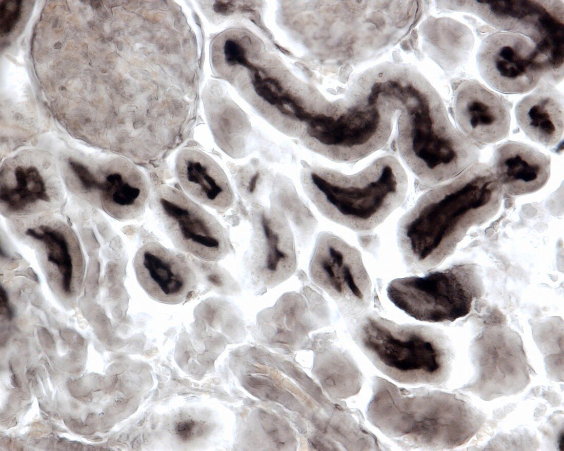
| Kidney proximal convoluted tubule, light micrograph. The main morphological difference between proximal and distal convoluted tubules is the presence in the proximal ones of a brush border made by densely packed microvilli, rich in alkaline phosphatase. In this image, a histochemical technique for alkaline phosphatase allows the identification of proximal convoluted tubules. Magnification: x360 when printed at 10 centimetres wide. | |
| Licence : | Droits gérés |
| Crédit: | Science Photo Library / JOSE CALVO |
| Taille de l’image : | 4674 px × 3739 px |
| Model Release : | Non requis |
| Property Release : | Non requis |
| Restrictions : | - |
Prix pour cette image À partir de 45 €
Produit vendu
(Calendrier, Carte postale, Carte de vœux, Impression sur textile, Packaging etc)
À partir de 45 €
Usage commercial
(Affichage, Annonce presse, Annonce TV, Carte, Digital - hors rés. sociaux, Digital - rés. sociaux etc)
À partir de 45 €
Éditorial
(Digital, Journal, Livre, Livre pratique, Magazine, Télévision etc)
À partir de 60 €
Usage non-commercial
(Digital - hors rés. sociaux, Digital - rés. sociaux etc)
À partir de 120 €
Mots clés
- anatomie,
- anatomique,
- biologie,
- biologique,
- canalicules,
- capillaire,
- capillaires,
- cellule,
- cellules,
- en bonne santé,
- histologie,
- histologique,
- micrographie optique,
- microscope,
- microscope optique,
- microscopie,
- microscopie optique,
- microscopique,
- néphron,
- néphrons,
- normal,
- rein,
- reins,
- sain,
- système urinaire,
- tissu conjonctif,
- tubulaire,
- tubule,
- tubule proximal compliqué,
- tubules,
- urinaire