Human small intestine, light micrograph
Numéro d’image : 12395454
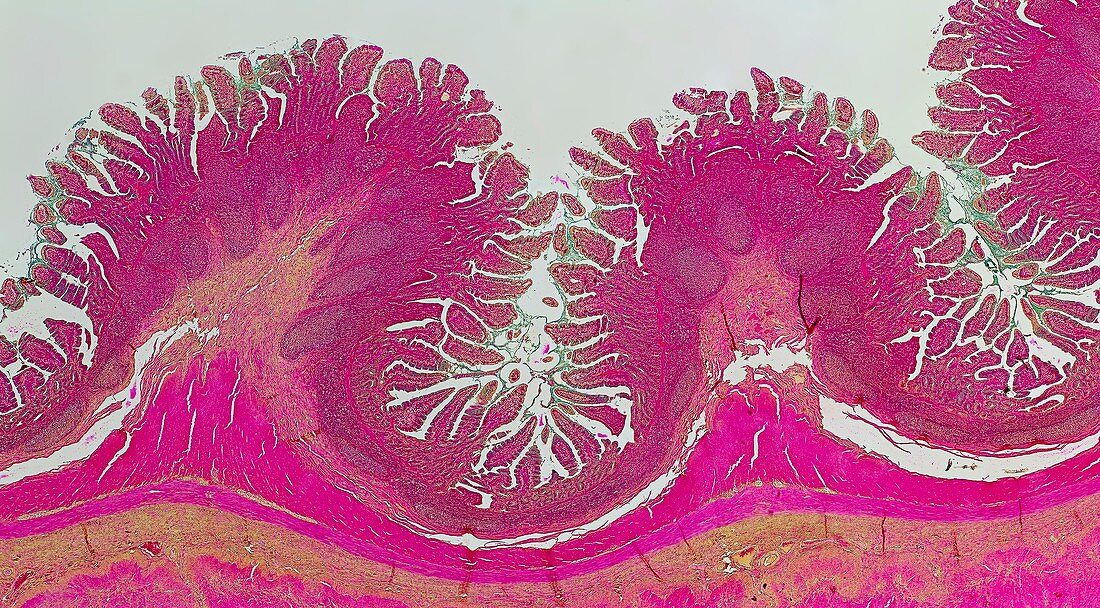
| Small intestine. Light micrograph of a transverse section through the human small intestine, showing its folded wall with villi. At top is the inner lumen of the intestine. At bottom are outer layers of muscles of the intestinal wall (pink). The inner intestinal wall contains folds, on which villi (purple) are seen. Villi are projections (0.5 to 1 millimetres long) which greatly increase the effective absorptive and secretory surface of the mucosa (mucus membrane) which lines the small intestine. The human small intestine is about 6 metres in length and requires a large surface area for the absorption of food which has been digested by enzymes. | |
| Licence : | Droits gérés |
| Crédit: | Science Photo Library / Fox, Frank |
| Taille de l’image : | 7952 px × 4396 px |
| Model Release : | Non requis |
| Property Release : | Non requis |
| Restrictions : | - |
Prix pour cette image À partir de 45 €
Produit vendu
(Calendrier, Carte postale, Carte de vœux, Impression sur textile, Packaging etc)
À partir de 45 €
Usage commercial
(Affichage, Annonce presse, Annonce TV, Carte, Digital - hors rés. sociaux, Digital - rés. sociaux etc)
À partir de 45 €
Éditorial
(Digital, Journal, Livre, Livre pratique, Magazine, Télévision etc)
À partir de 60 €
Usage non-commercial
(Digital - hors rés. sociaux, Digital - rés. sociaux etc)
À partir de 120 €