Skeletal bone core surface, SEM
Numéro d’image : 12377231
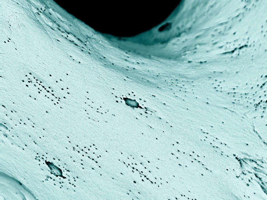
| Skeletal bone core surface. Coloured scanning electron micrograph (SEM) of the surface of bone tissue in a bone core taken from a femur (thigh bone) and cleaned with enzymes to remove all soft tissues. This reveals the underlying bone structure, which includes osteocytes (bone cells) in this close-up view. Imaged using a backscatter electron detector (BSD). | |
| Licence : | Droits gérés |
| Crédit: | Science Photo Library / UNIVERSITY OF ABERDEEN / Kevin Mackenzie |
| Taille de l’image : | 3413 px × 2559 px |
| Model Release : | Non requis |
| Property Release : | Non requis |
| Restrictions : | - |
Prix pour cette image À partir de 45 €
Produit vendu
(Calendrier, Carte postale, Carte de vœux, Impression sur textile, Packaging etc)
À partir de 45 €
Usage commercial
(Affichage, Annonce presse, Annonce TV, Carte, Digital - hors rés. sociaux, Digital - rés. sociaux etc)
À partir de 45 €
Éditorial
(Digital, Journal, Livre, Livre pratique, Magazine, Télévision etc)
À partir de 60 €
Usage non-commercial
(Digital - hors rés. sociaux, Digital - rés. sociaux etc)
À partir de 120 €
Mots clés
- anatomie,
- anatomique,
- aucun,
- biologie,
- biologique,
- coloré,
- colorié,
- colorisé,
- corps humain,
- détail,
- en bonne santé,
- fémur,
- gros plan,
- M.E.B.,
- MEB,
- microscope électronique à balayage,
- minéral,
- minérale,
- minéraux,
- normal,
- os,
- os de la cuisse,
- os du squelette,
- osseuse,
- osseux,
- ostéocyte,
- ostéologie,
- personne,
- sain,
- surface,
- tissus,
- trabecula,
- trabeculae,
- trabécule,
- trabéculum