Secretory cell desmosomes, TEM
Numéro d’image : 12360631
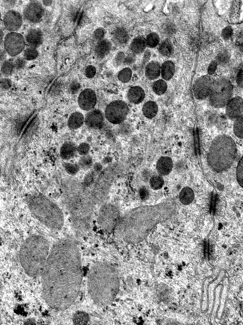
| Transmission electron micrograph (TEM) showing the cytoplasm of secretory cells. In the top of the image appear secretory granules (very dark). In the bottom half, several mitochondria can be seen. At the intercellular borders several desmosomes (junctions) stand out. | |
| Licence : | Droits gérés |
| Crédit: | Science Photo Library / JOSE CALVO |
| Taille de l’image : | 3617 px × 4841 px |
| Model Release : | Non requis |
| Property Release : | Non requis |
| Restrictions : | - |
Prix pour cette image À partir de 45 €
Produit vendu
(Calendrier, Carte postale, Carte de vœux, Impression sur textile, Packaging etc)
À partir de 45 €
Usage commercial
(Affichage, Annonce presse, Annonce TV, Carte, Digital - hors rés. sociaux, Digital - rés. sociaux etc)
À partir de 45 €
Éditorial
(Digital, Journal, Livre, Livre pratique, Magazine, Télévision etc)
À partir de 60 €
Usage non-commercial
(Digital - hors rés. sociaux, Digital - rés. sociaux etc)
À partir de 120 €
Mots clés
- biologie,
- biologique,
- cellule,
- céllule sécrétrice,
- colorisé,
- cytologie,
- cytologique,
- cytoplasme,
- desmosome,
- granules,
- granulés,
- histologie,
- histologique,
- M.E.T.,
- membrane,
- MET,
- micrographie,
- micrographie électronique à transmission,
- microscope,
- microscope électronique,
- microscope électronique à transmission,
- microscopie,
- mitochondria,
- mitochondrie,
- mitochondries,
- ultrastructure,
- ultrastructure des cellules