Pine needle, light micrograph
Numéro d’image : 12324906
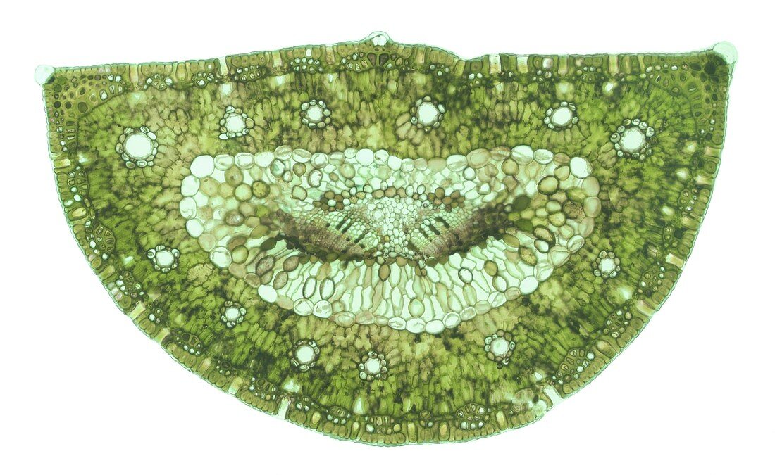
| Light micrograph of a cross section through a pine (Pinus subgenus Pinus) needle, or leaf. The outer epidermal layer is covered by a thin cuticle (pale blue). Regularly spaced stomata are present on both the upper and lower surfaces of the leaf. A ring of 12 evenly spaced resin canals are located within the mesophyll layer. A layer of endodermis surrounds transfusional tissue, which in turn contains two vascular bundles composed of phloem (lower layer of smaller cells) and xylem (upper layer). Magnification: x40 at 10cm wide. | |
| Licence : | Droits gérés |
| Crédit: | Science Photo Library / Lowry, Steve |
| Taille de l’image : | 6289 px × 3862 px |
| Model Release : | Non requis |
| Property Release : | Non requis |
| Restrictions : | - |
Prix pour cette image À partir de 45 €
Produit vendu
(Calendrier, Carte postale, Carte de vœux, Impression sur textile, Packaging etc)
À partir de 45 €
Usage commercial
(Affichage, Annonce presse, Annonce TV, Carte, Digital - hors rés. sociaux, Digital - rés. sociaux etc)
À partir de 45 €
Éditorial
(Digital, Journal, Livre, Livre pratique, Magazine, Télévision etc)
À partir de 60 €
Usage non-commercial
(Digital - hors rés. sociaux, Digital - rés. sociaux etc)
À partir de 120 €