SEM of epithelium in the oesophagus
Numéro d’image : 12304379
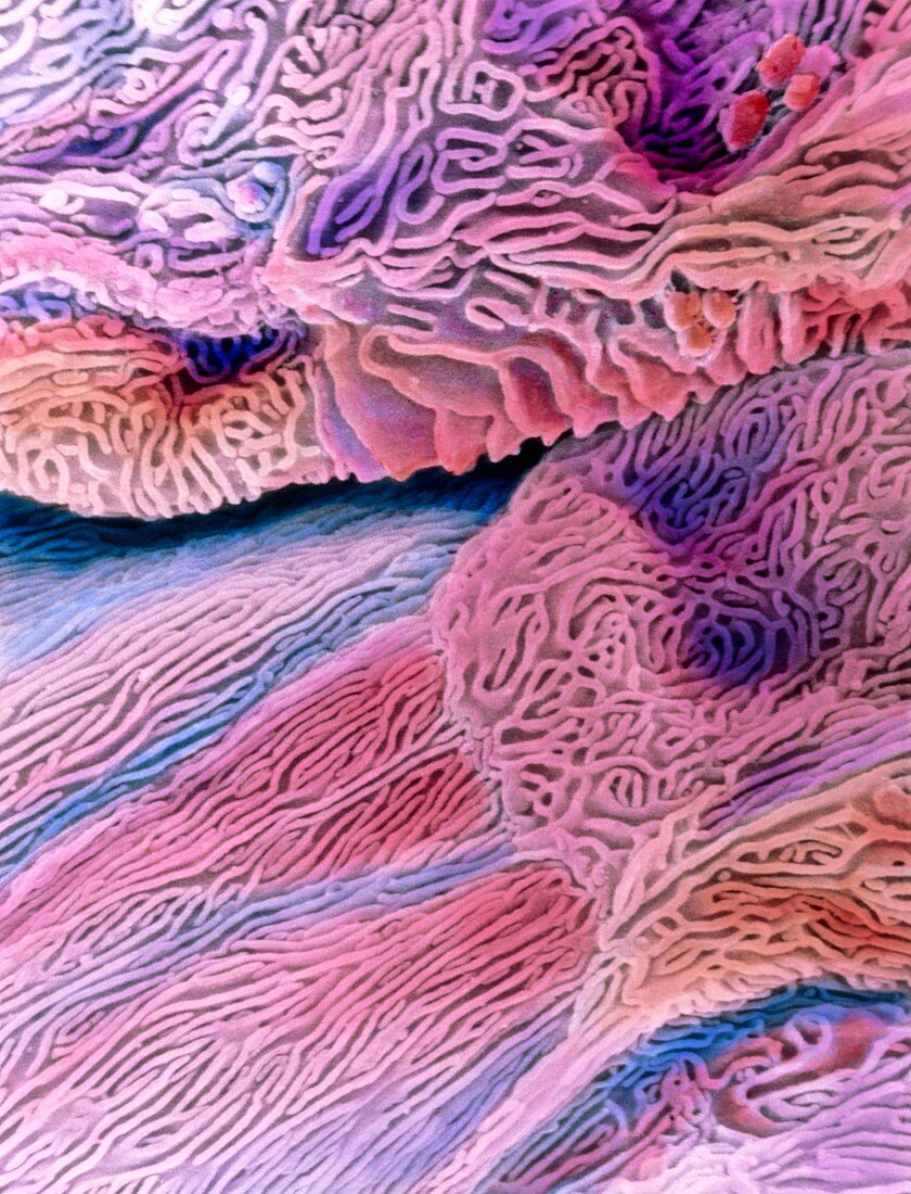
| False-colour scanning electron micrograph (SEM) of the epithelium in the oesophagus. Individual epithelial cells are seen here, each with a highly folded surface. These microfolds are called microplicae. At centre right, an obvious boundary to one cell can be seen. This stratified squamous epithelium consists of flattened cells that occur many layers thick. As a muscular non-absorptive tube, the oesophagus transports swallowed food to the stomach. The microfolds on the cells keep the oesophagus moist; trap mucous to lubricate passing food; and strengthen the epithelium against food abrasion. Magnification: x1, 950 at 6x7cm size. Magnification: x3, 125 at 4x5 inch size. | |
| Licence : | Droits gérés |
| Crédit: | Science Photo Library / DEPT. OF ANATOMY / PROF. P. MOTTA |
| Taille de l’image : | 3692 px × 4836 px |
| Model Release : | Non requis |
| Property Release : | Non requis |
| Restrictions : | - |
Prix pour cette image À partir de 45 €
Produit vendu
(Calendrier, Carte postale, Carte de vœux, Impression sur textile, Packaging etc)
À partir de 45 €
Usage commercial
(Affichage, Annonce presse, Annonce TV, Carte, Digital - hors rés. sociaux, Digital - rés. sociaux etc)
À partir de 45 €
Éditorial
(Digital, Journal, Livre, Livre pratique, Magazine, Télévision etc)
À partir de 60 €
Usage non-commercial
(Digital - hors rés. sociaux, Digital - rés. sociaux etc)
À partir de 120 €