Goblet cell in the lining of the nasal epithelium, TEM
Numéro d’image : 12297996
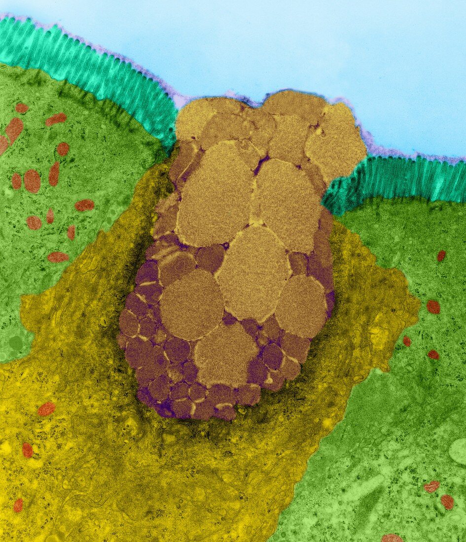
| Goblet cell in the lining of the nasal epithelium, coloured transmission electron micrograph (TEM). The goblet cell (yellow) is full of mucus granules (brown) and is surrounded by microvilli (turquoise) coated columnar epithelial cells (light green). The squamous nasal epithelium is made up of both ciliated (not seen) and microvilli coated columnar epithelial cells. Goblet cells are interspersed through this nasal epithelial layer. The goblet cells secrete mucus onto the surface of the nasal cavity (where the surface of the ciliated and microvilli coated columnar epithelial cells are exposed in the nasal cavity). Cilia and microvilli become coated with sticky mucus that helps trap foreign objects, such as dust and pathogens, preventing them from entering the lungs. Coordinated, wave-like beating of the cilia propels the mucus to the back of the nasopharynx, where it is swallowed, thus removing foreign objects. Magnification: x2, 030 when shortest axis | |
| Licence : | Droits gérés |
| Crédit: | Science Photo Library / DENNIS KUNKEL MICROSCOPY |
| Taille de l’image : | 2740 px × 3190 px |
| Model Release : | Non requis |
| Property Release : | Non requis |
| Restrictions : | - |
Prix pour cette image À partir de 45 €
Produit vendu
(Calendrier, Carte postale, Carte de vœux, Impression sur textile, Packaging etc)
À partir de 45 €
Usage commercial
(Affichage, Annonce presse, Annonce TV, Carte, Digital - hors rés. sociaux, Digital - rés. sociaux etc)
À partir de 45 €
Éditorial
(Digital, Journal, Livre, Livre pratique, Magazine, Télévision etc)
À partir de 60 €
Usage non-commercial
(Digital - hors rés. sociaux, Digital - rés. sociaux etc)
À partir de 120 €