Fluorescence mapping of an oncogene
Numéro d’image : 12248853
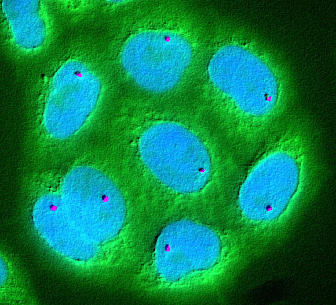
| Fluorescence mapping of an oncogene. Fluorescence light micrograph showing a close-up of cell nuclei, mapping the position of genes. This research aids understanding of what governs the genome. Here, a single gene called PEM (polymorphic epithelial mucin, purple) has been localized using fluorescence in-situ hybridization. DNA is stained blue, while the cell cytoplasm is stained green. Overexpression of PEM (also called MUC1) is often associated with colon, breast, ovarian, lung and pancreatic cancers. An oncogene is a gene that causes cancer. | |
| Licence : | Droits gérés |
| Crédit: | Science Photo Library / NATIONAL CANCER INSTITUTE / NCI Center for Cancer Research |
| Taille de l’image : | 3117 px × 2824 px |
| Model Release : | Non requis |
| Property Release : | Non requis |
| Restrictions : |
|
Prix pour cette image À partir de 45 €
Produit vendu
(Calendrier, Carte postale, Carte de vœux, Impression sur textile, Packaging etc)
À partir de 45 €
Usage commercial
(Affichage, Annonce presse, Annonce TV, Carte, Digital - hors rés. sociaux, Digital - rés. sociaux etc)
À partir de 45 €
Éditorial
(Digital, Journal, Livre, Livre pratique, Magazine, Télévision etc)
À partir de 60 €
Usage non-commercial
(Digital - hors rés. sociaux, Digital - rés. sociaux etc)
À partir de 120 €
Mots clés
- anormal,
- aucun,
- cancer,
- cancéreux,
- cellulaire,
- cellule,
- cellule cancéreuse,
- cellules,
- désordre,
- état,
- fluorescence,
- fluorescent,
- gène,
- génétique,
- maladie,
- malignité,
- malin,
- malsain,
- médecine,
- médical,
- médicale,
- micrographie optique,
- microscope optique,
- microscopie optique,
- noyau,
- noyeaux cellulaires,
- noyeaux de cellules,
- nucleus,
- oncogène,
- oncologie,
- personne,
- recherche,
- tissus,
- trouble,
- tumeur,
- tumeur maligne