X-ray of an elderly woman's fractured fibula bone
Numéro d’image : 12100144
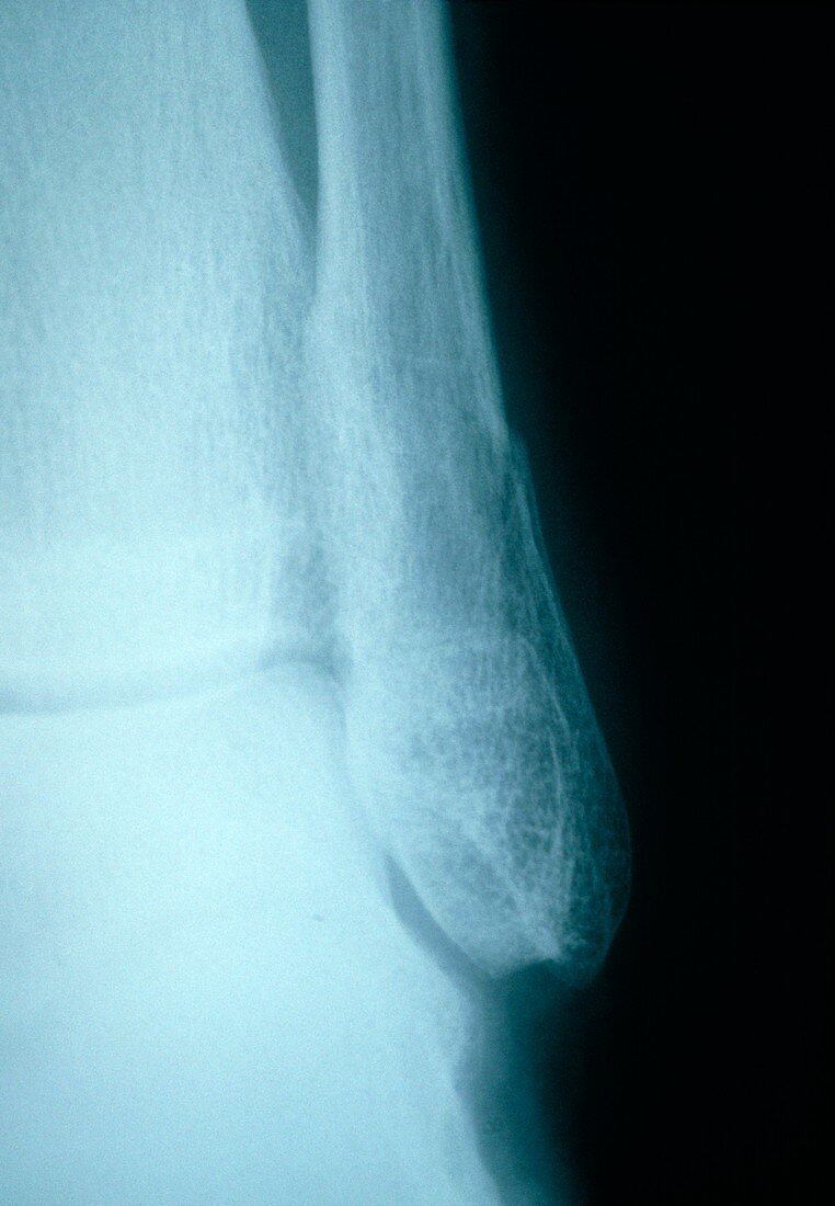
| Broken ankle. X-ray of the ankle joint of a 60 year old woman showing a fractured fibula. The fibula is the long,thin bone descending from top centre. The fracture appears as a dark crevice just above centre on the right edge of the fibula. To the left of the fibula is the tibia (shin bone),with the bones of the foot at bottom left. Broken bones are fairly common among elderly women as their bones become brittle after the menopause due to a lack of the hormone oestrogen. Fractures such as this cause pain and severely limit joint movement,and may also be associated with torn ligaments in the area. A fracture such as this has to be set in a cast,and may take months to heal | |
| Licence : | Droits gérés |
| Crédit: | Science Photo Library / Marazzi, Dr. P. |
| Taille de l’image : | 3520 px × 5079 px |
| Model Release : | Non requis |
| Property Release : | Non requis |
| Restrictions : | - |
Prix pour cette image À partir de 45 €
Produit vendu
(Calendrier, Carte postale, Carte de vœux, Impression sur textile, Packaging etc)
À partir de 45 €
Usage commercial
(Affichage, Annonce presse, Annonce TV, Carte, Digital - hors rés. sociaux, Digital - rés. sociaux etc)
À partir de 45 €
Éditorial
(Digital, Journal, Livre, Livre pratique, Magazine, Télévision etc)
À partir de 60 €
Usage non-commercial
(Digital - hors rés. sociaux, Digital - rés. sociaux etc)
À partir de 120 €