Developing nail,LM
Numéro d’image : 12099973
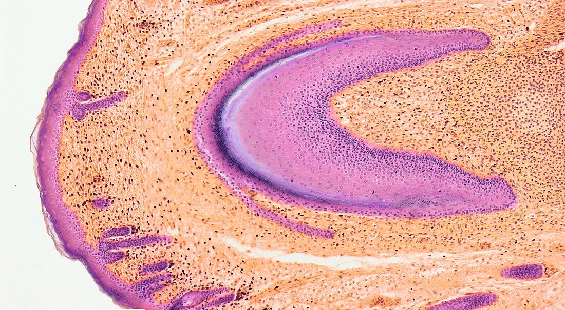
| Developing nail. Light micrograph (LM) of longitudinal section through a fetal finger tip to show the developing nail. The large area of pink nail bed epithelium is tipped by the developing nail. Orange connective tissue (dermis) forms the bulk of the image. Pink stratified squamous epithelium of the skin is far left. Magnification: x15 when printed at 10 centimetres wide | |
| Licence : | Droits gérés |
| Crédit: | Science Photo Library / Gschmeissner, Steve |
| Taille de l’image : | 6096 px × 3352 px |
| Model Release : | Non requis |
| Property Release : | Non requis |
| Restrictions : | - |
Prix pour cette image À partir de 45 €
Produit vendu
(Calendrier, Carte postale, Carte de vœux, Impression sur textile, Packaging etc)
À partir de 45 €
Usage commercial
(Affichage, Annonce presse, Annonce TV, Carte, Digital - hors rés. sociaux, Digital - rés. sociaux etc)
À partir de 45 €
Éditorial
(Digital, Journal, Livre, Livre pratique, Magazine, Télévision etc)
À partir de 60 €
Usage non-commercial
(Digital - hors rés. sociaux, Digital - rés. sociaux etc)
À partir de 120 €
Mots clés
- biologie,
- biologique,
- bout de doigt,
- bout des doigts,
- bout du doigt,
- catégorie,
- corps humain,
- coupe,
- divisé,
- en bonne santé,
- épithélium squameux stratifié,
- foetal,
- foetale,
- fœtale,
- foetus,
- histologie,
- histologique,
- lit d'ongle,
- micrographie optique,
- microscope optique,
- microscopie optique,
- normal,
- ongle,
- partie,
- peau,
- sain,
- section,
- tissu adipeux