Foetal finger
Numéro d’image : 12071261
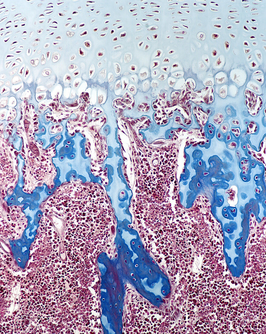
| Light micrograph of a developing finger bone in a late foetus. This micrograph shows hypertrophying cartilage and mixed bone spicules within the marrow space. The spicules contain an inner core of calcified cartilage (stained pale blue) and bone tissue (stained dark blue) deposited against the calcified cartilage. Mallory-Azan stain. Magnification: x175 | |
| Licence : | Droits gérés |
| Crédit: | Science Photo Library / Ross, Michael |
| Taille de l’image : | 1500 px × 1880 px |
| Model Release : | Non requis |
| Property Release : | Non requis |
| Restrictions : |
|
Prix pour cette image À partir de 45 €
Produit vendu
(Calendrier, Carte postale, Carte de vœux, Impression sur textile, Packaging etc)
À partir de 45 €
Usage commercial
(Affichage, Annonce presse, Annonce TV, Carte, Digital - hors rés. sociaux, Digital - rés. sociaux etc)
À partir de 45 €
Éditorial
(Digital, Journal, Livre, Livre pratique, Magazine, Télévision etc)
À partir de 60 €
Usage non-commercial
(Digital - hors rés. sociaux, Digital - rés. sociaux etc)
À partir de 120 €
Mots clés
- anatomie,
- corps humain,
- développement,
- developpement des os,
- développement du squelette,
- développement foetal,
- développment osseux,
- doigts,
- épiphyse,
- foetal,
- foetale,
- foetus,
- histologie,
- micrographie,
- microscope optique,
- microscopie,
- microscopie optique,
- os,
- phalange,
- spicule,
- spicules,
- tissu osseux