Foetal finger
Numéro d’image : 12071259
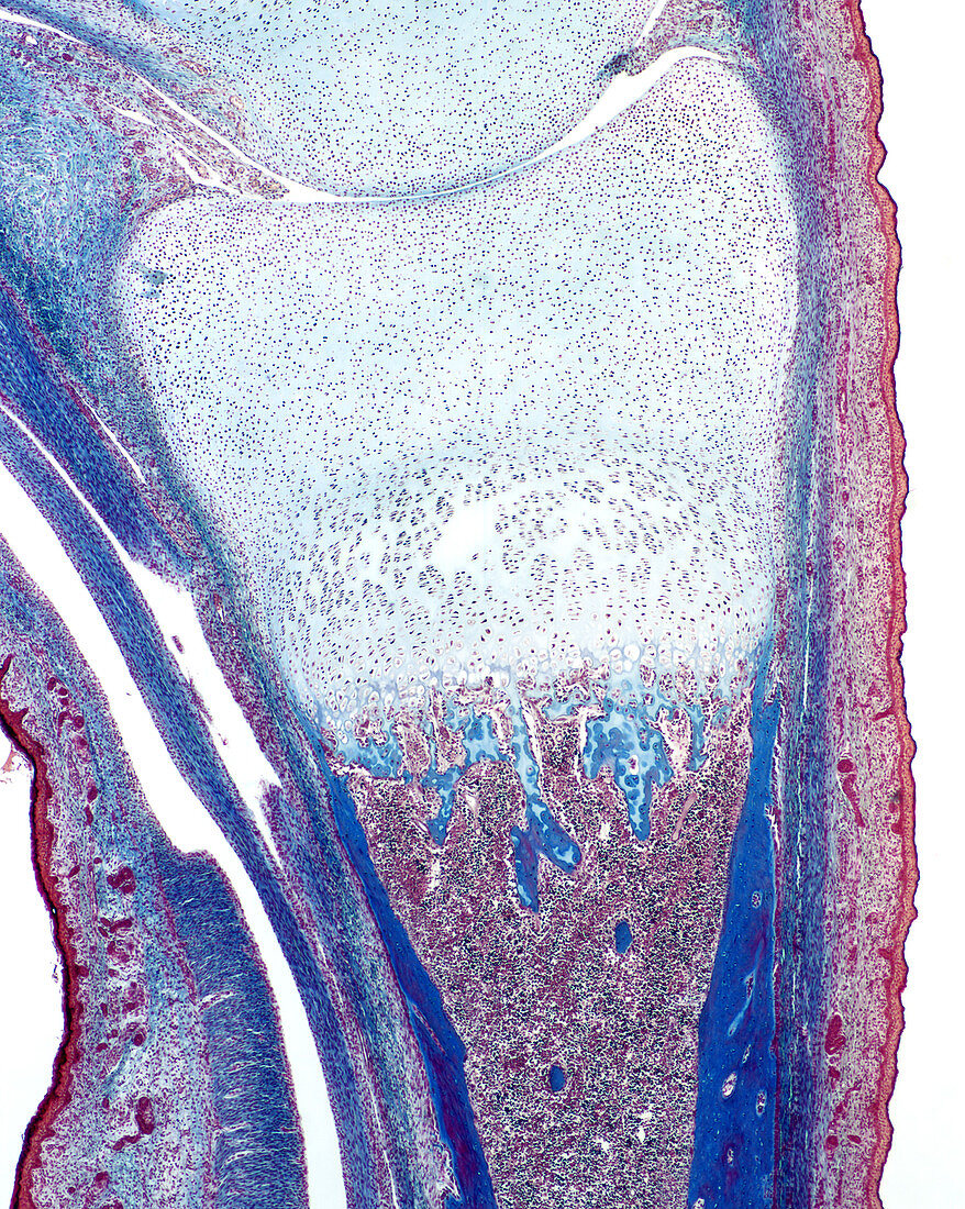
| Light micrograph of a developing finger bone in a late foetus. At the top is a joint cavity where the medial phalange is articulating with the proximal phalange. The heads of the two long bones,the epiphyses,consist entirely of cartilage at this stage of development. Mixed spicules consisting of bone and cartilage are seen in the marrow portion of the bone. Bone tissue,stained dark blue,is also present in the diaphysis of the bone. Mallory-Azan stain. Magnification: x45 | |
| Licence : | Droits gérés |
| Crédit: | Science Photo Library / Ross, Michael |
| Taille de l’image : | 1500 px × 1880 px |
| Model Release : | Non requis |
| Property Release : | Non requis |
| Restrictions : |
|
Prix pour cette image À partir de 45 €
Produit vendu
(Calendrier, Carte postale, Carte de vœux, Impression sur textile, Packaging etc)
À partir de 45 €
Usage commercial
(Affichage, Annonce presse, Annonce TV, Carte, Digital - hors rés. sociaux, Digital - rés. sociaux etc)
À partir de 45 €
Éditorial
(Digital, Journal, Livre, Livre pratique, Magazine, Télévision etc)
À partir de 60 €
Usage non-commercial
(Digital - hors rés. sociaux, Digital - rés. sociaux etc)
À partir de 120 €
Mots clés
- anatomie,
- cartilage,
- corps humain,
- croissance osseuse,
- développement,
- developpement des os,
- développement du squelette,
- développement foetal,
- développment osseux,
- doigt,
- doigts,
- foetal,
- foetale,
- foetus,
- histologie,
- micrographe,
- micrographes optiques,
- micrographie,
- microscope,
- microscope optique,
- microscopie,
- microscopie optique,
- os,
- phalange,
- phalanges,
- tissu osseux