56-day-old Embryo (Micro-MRI)
Numéro d’image : 12071245
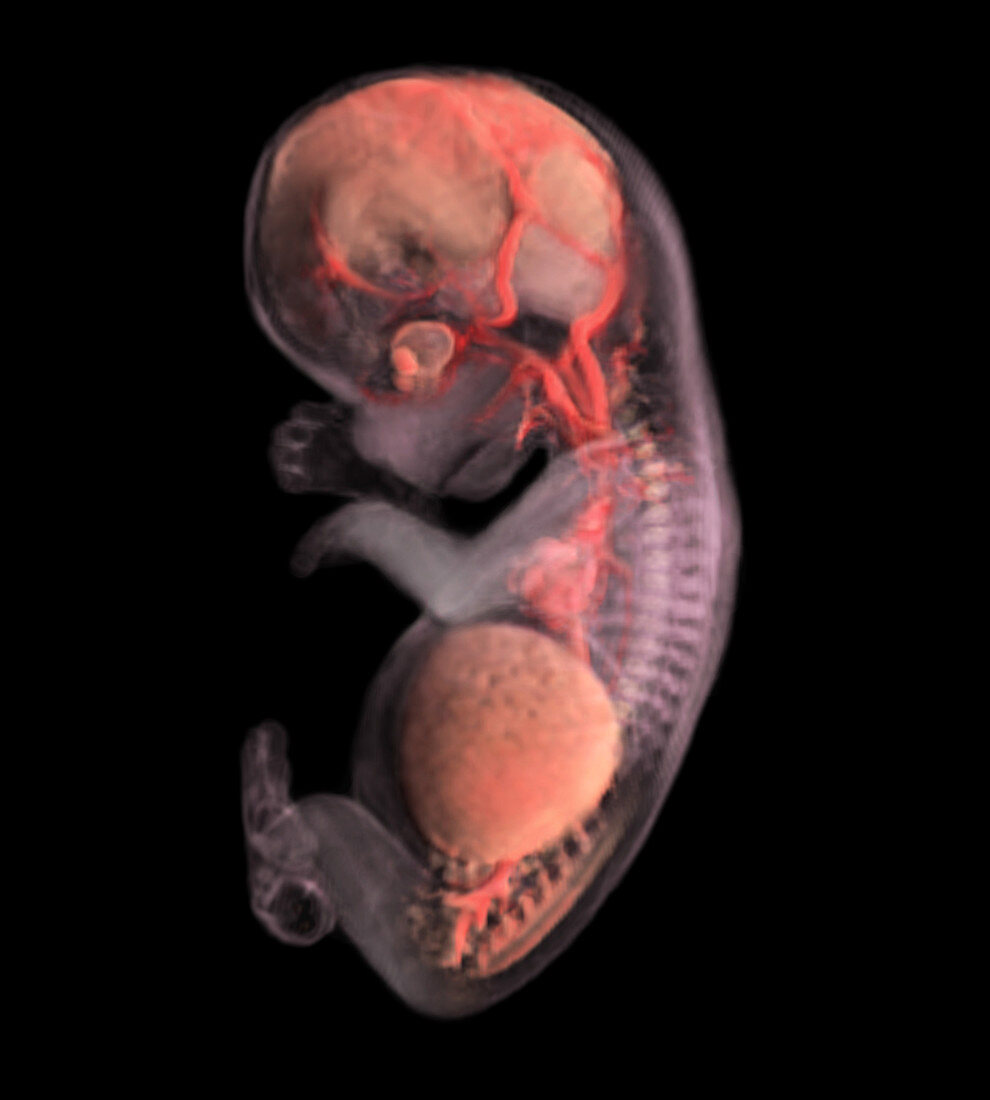
| Computer generated image reconstructed from Micro-MRI,actual size of embryo = 30.0 mm - This left-sided image of the embryo at eight weeks of development has been altered so that the skin has a whitish-translucent appearance and the major organs are highlighted in pink,with the major blood vessels emphasized in red. The age is calculated by the day of fertilization. The image also provides the opportunity to view the internal organs of the embryo within the context of its body form. It can be seen from this image that the brain in the head region and the liver in the lower part of the body occupy the greatest portion embryos body mass. The skeletal structure is slightly hinted in white,as the image reveals the ridges of the vertebrae and outlines of the fingers and toes. Image from the book From Conception to Birth: A Life Unfolds | |
| Licence : | Droits gérés |
| Crédit: | Science Photo Library / Anatomical Travelogue |
| Taille de l’image : | 2767 px × 3075 px |
| Model Release : | Non requis |
| Property Release : | Non requis |
| Restrictions : |
|
Prix pour cette image À partir de 45 €
Produit vendu
(Calendrier, Carte postale, Carte de vœux, Impression sur textile, Packaging etc)
À partir de 45 €
Usage commercial
(Affichage, Annonce presse, Annonce TV, Carte, Digital - hors rés. sociaux, Digital - rés. sociaux etc)
À partir de 45 €
Éditorial
(Digital, Journal, Livre, Livre pratique, Magazine, Télévision etc)
À partir de 60 €
Usage non-commercial
(Digital - hors rés. sociaux, Digital - rés. sociaux etc)
À partir de 120 €