44-day-old Embryo (Micro-MRI)
Numéro d’image : 12071243
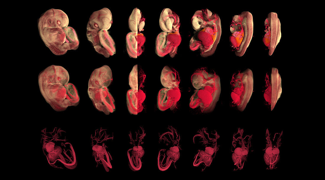
| Computer generated image reconstructed from Micro-MRI,actual size of embryo = 13.0 mm - This sequential series of images unravel the structural make-up of a human embryo,from the outer appearance to revealing the intricate components inside. The embryo focused here is in the seventh week of development. Age is calculated from the day of fertilization. These 360 degree rotations show the embryo with a half split connective tissue (upper row),half split connective tissue with circulatory system (middle row) and circulatory system only (lower row ). Image from the book From Conception to Birth: A Life Unfolds | |
| Licence : | Droits gérés |
| Crédit: | Science Photo Library / Anatomical Travelogue |
| Taille de l’image : | 6674 px × 3708 px |
| Model Release : | Non requis |
| Property Release : | Non requis |
| Restrictions : |
|
Prix pour cette image À partir de 45 €
Produit vendu
(Calendrier, Carte postale, Carte de vœux, Impression sur textile, Packaging etc)
À partir de 45 €
Usage commercial
(Affichage, Annonce presse, Annonce TV, Carte, Digital - hors rés. sociaux, Digital - rés. sociaux etc)
À partir de 45 €
Éditorial
(Digital, Journal, Livre, Livre pratique, Magazine, Télévision etc)
À partir de 60 €
Usage non-commercial
(Digital - hors rés. sociaux, Digital - rés. sociaux etc)
À partir de 120 €