28-day-old Embryo (Micro-MRI)
Numéro d’image : 12071239
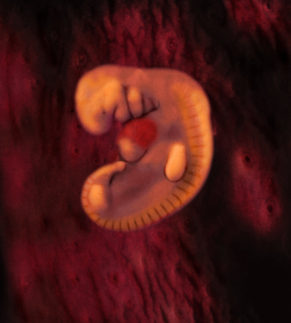
| Micro-MRI,reconstructed with 3D imagery,actual size of embryo = 4.0 mm - The image depicts a human embryo during its fourth week of development. Age is calculated from the day of fertalization. In this image,the fusing tubes of the heart is highlighted in red. Early growth of the cardiovascular system begins during the third week,when blood vessels form,and continue into the following weeks of development. As the heart grows and elongates,it will start to form an S-shape and establish divisions within itself. The region of the back highlighted in yellow is pairs of somites. The somites will lead to the development of skeletal structures and the dermis of the skin. (left view) Image from the book From Conception to Birth: A Life Unfolds | |
| Licence : | Droits gérés |
| Crédit: | Science Photo Library / Anatomical Travelogue |
| Taille de l’image : | 2775 px × 3083 px |
| Model Release : | Non requis |
| Property Release : | Non requis |
| Restrictions : |
|
Prix pour cette image À partir de 45 €
Produit vendu
(Calendrier, Carte postale, Carte de vœux, Impression sur textile, Packaging etc)
À partir de 45 €
Usage commercial
(Affichage, Annonce presse, Annonce TV, Carte, Digital - hors rés. sociaux, Digital - rés. sociaux etc)
À partir de 45 €
Éditorial
(Digital, Journal, Livre, Livre pratique, Magazine, Télévision etc)
À partir de 60 €
Usage non-commercial
(Digital - hors rés. sociaux, Digital - rés. sociaux etc)
À partir de 120 €