TEM of metaphase cell division of a HeLa cell
Numéro d’image : 12071186
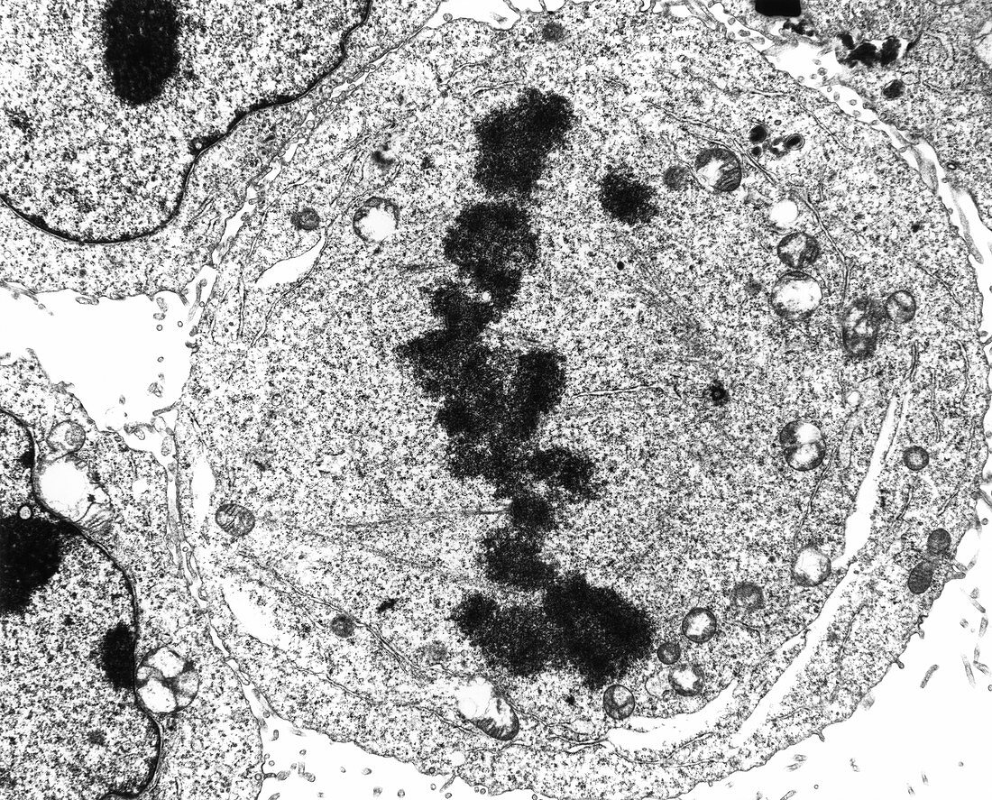
| Metaphase cell division. Transmission Electron Micrograph (TEM) of a section through a HeLa cell during metaphase cell division (mitosis). At centre are chromosomes (dark structures); during metaphase the chromosomes line up in one plane in the centre of the cell,in preparation for nuclear division. The chromosomes are moved by spindle microtubules,fine straight lines leading to a centriole (dark circle,centre right). Organelles (white) are visible in the cytoplasm. During mitosis a cell produces two genetically identical daughter cells. HeLa cells are human cancer cells cultured in the laboratory and used in cancer research. Magnification: x9,175 at 8x10 inch size | |
| Licence : | Droits gérés |
| Crédit: | Science Photo Library / Murti, Dr. Gopal |
| Taille de l’image : | 4706 px × 3796 px |
| Model Release : | Non requis |
| Property Release : | Non requis |
| Restrictions : | - |
Prix pour cette image À partir de 45 €
Produit vendu
(Calendrier, Carte postale, Carte de vœux, Impression sur textile, Packaging etc)
À partir de 45 €
Usage commercial
(Affichage, Annonce presse, Annonce TV, Carte, Digital - hors rés. sociaux, Digital - rés. sociaux etc)
À partir de 45 €
Éditorial
(Digital, Journal, Livre, Livre pratique, Magazine, Télévision etc)
À partir de 60 €
Usage non-commercial
(Digital - hors rés. sociaux, Digital - rés. sociaux etc)
À partir de 120 €