Confocal optical section of mouse kidney,
Numéro d’image : 12070898
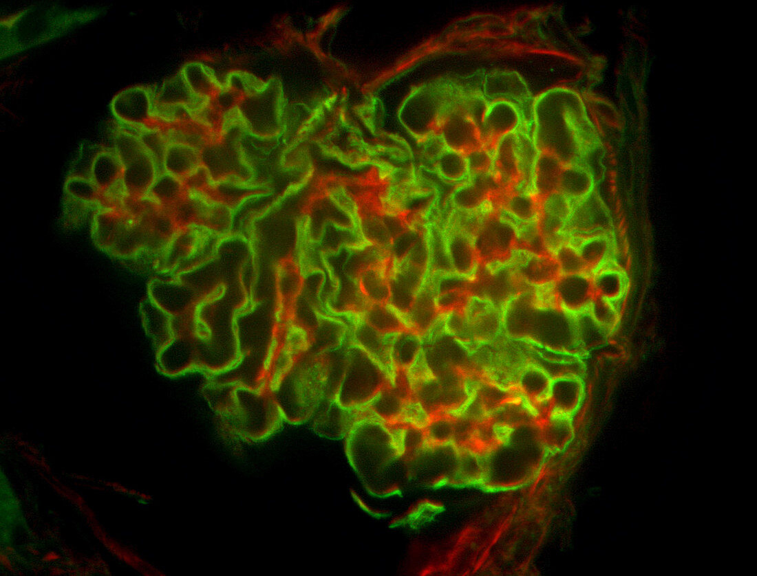
| A spinning disk confocal optical section of mouse kidney stained for WGA (green) labeling glycoproteins in the cell membrane, and actin (red). The image of the living cell is gained through a fast acquisition confocal microscope. | |
| Licence : | Droits gérés |
| Crédit: | Science Photo Library / Waters, Jennifer |
| Taille de l’image : | 3608 px × 2749 px |
| Model Release : | Non requis |
| Property Release : | Non requis |
| Restrictions : |
|
Prix pour cette image À partir de 45 €
Produit vendu
(Calendrier, Carte postale, Carte de vœux, Impression sur textile, Packaging etc)
À partir de 45 €
Usage commercial
(Affichage, Annonce presse, Annonce TV, Carte, Digital - hors rés. sociaux, Digital - rés. sociaux etc)
À partir de 45 €
Éditorial
(Digital, Journal, Livre, Livre pratique, Magazine, Télévision etc)
À partir de 60 €
Usage non-commercial
(Digital - hors rés. sociaux, Digital - rés. sociaux etc)
À partir de 120 €