colourized Frontal X-ray from an IVP Version 2
Numéro d’image : 12070894
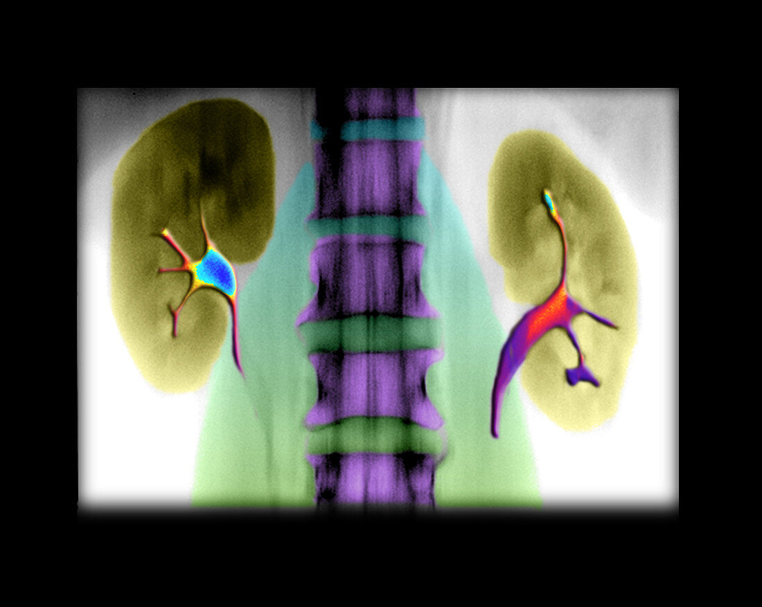
| colour enhanced x-ray showing a frontal view from an IVP (intravenous pyelogram). An IVP is an enhanced x-ray image in which a contrast material (dye) is used to examine the anatomy and function of the kidneys,ureters,and urinary bladder. The kidneys are on either side of the spine in the middle of the image. They connect to the urinary bladder via the ureters which are the skinny little tubes running vertically in this image | |
| Licence : | Droits gérés |
| Crédit: | Science Photo Library / Living Art Enterprises, LLC |
| Taille de l’image : | 3762 px × 3000 px |
| Model Release : | Non requis |
| Property Release : | Non requis |
| Restrictions : |
|
Prix pour cette image À partir de 45 €
Produit vendu
(Calendrier, Carte postale, Carte de vœux, Impression sur textile, Packaging etc)
À partir de 45 €
Usage commercial
(Affichage, Annonce presse, Annonce TV, Carte, Digital - hors rés. sociaux, Digital - rés. sociaux etc)
À partir de 45 €
Éditorial
(Digital, Journal, Livre, Livre pratique, Magazine, Télévision etc)
À partir de 60 €
Usage non-commercial
(Digital - hors rés. sociaux, Digital - rés. sociaux etc)
À partir de 120 €