LM of a villus in the small intestine
Numéro d’image : 12070830
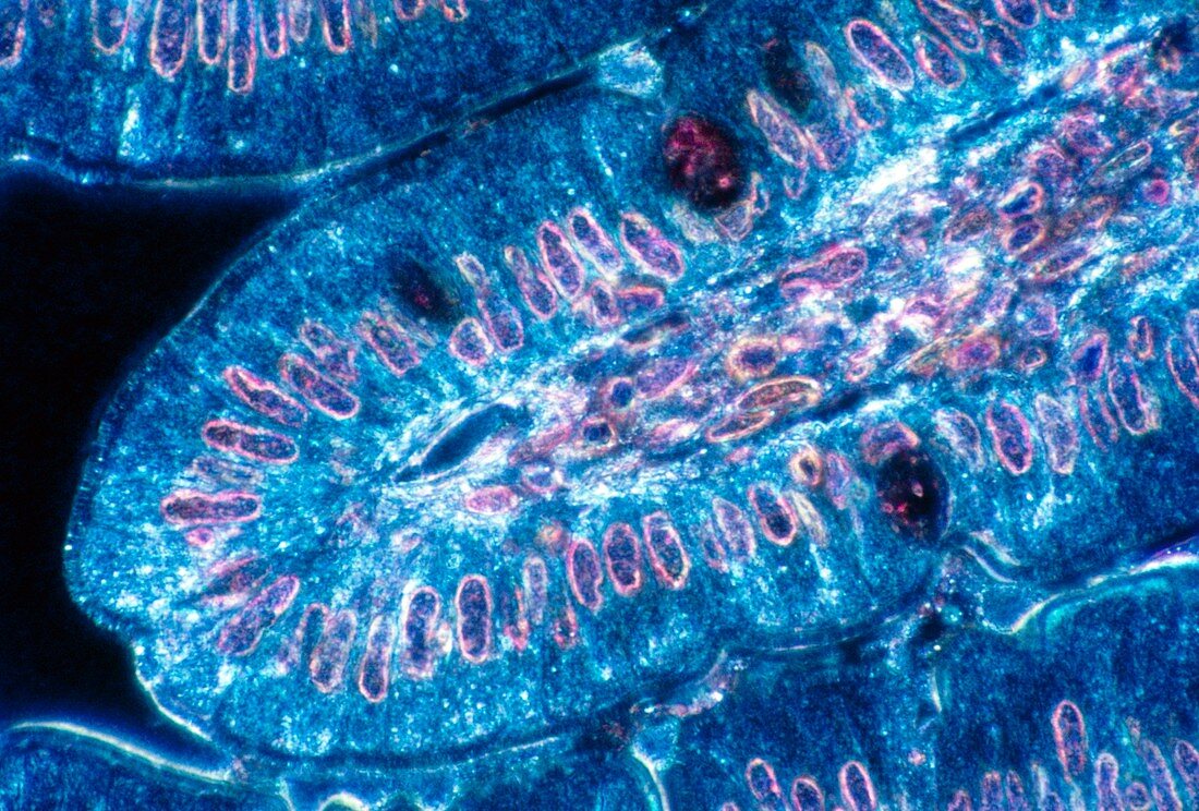
| Intestinal villus. Light micrograph of a section through a villus in the small intestine. Villi are projections on the intestinal wall adapted for food absorption. At centre,the core of the villus contains blood capillaries. The exterior wall is lined by columnar epithelial cells,each with a nucleus (pink/purple). These cells are specialised for food absorption,assisted by a dark blue "brush border" on their external surface. Food then passes into the bloodstream. Scattered among the epithelial cells are goblet cells (large,dark-staining) which secrete mucous to assist the movement of food through the gut. Toluidine blue stain. Magnification: x800 at 35mm size | |
| Licence : | Droits gérés |
| Crédit: | Science Photo Library / Fox, Cecil H. |
| Taille de l’image : | 5092 px × 3445 px |
| Model Release : | Non requis |
| Property Release : | Non requis |
| Restrictions : |
|
Prix pour cette image À partir de 45 €
Produit vendu
(Calendrier, Carte postale, Carte de vœux, Impression sur textile, Packaging etc)
À partir de 45 €
Usage commercial
(Affichage, Annonce presse, Annonce TV, Carte, Digital - hors rés. sociaux, Digital - rés. sociaux etc)
À partir de 45 €
Éditorial
(Digital, Journal, Livre, Livre pratique, Magazine, Télévision etc)
À partir de 60 €
Usage non-commercial
(Digital - hors rés. sociaux, Digital - rés. sociaux etc)
À partir de 120 €