Epigastric region
Numéro d’image : 12070816
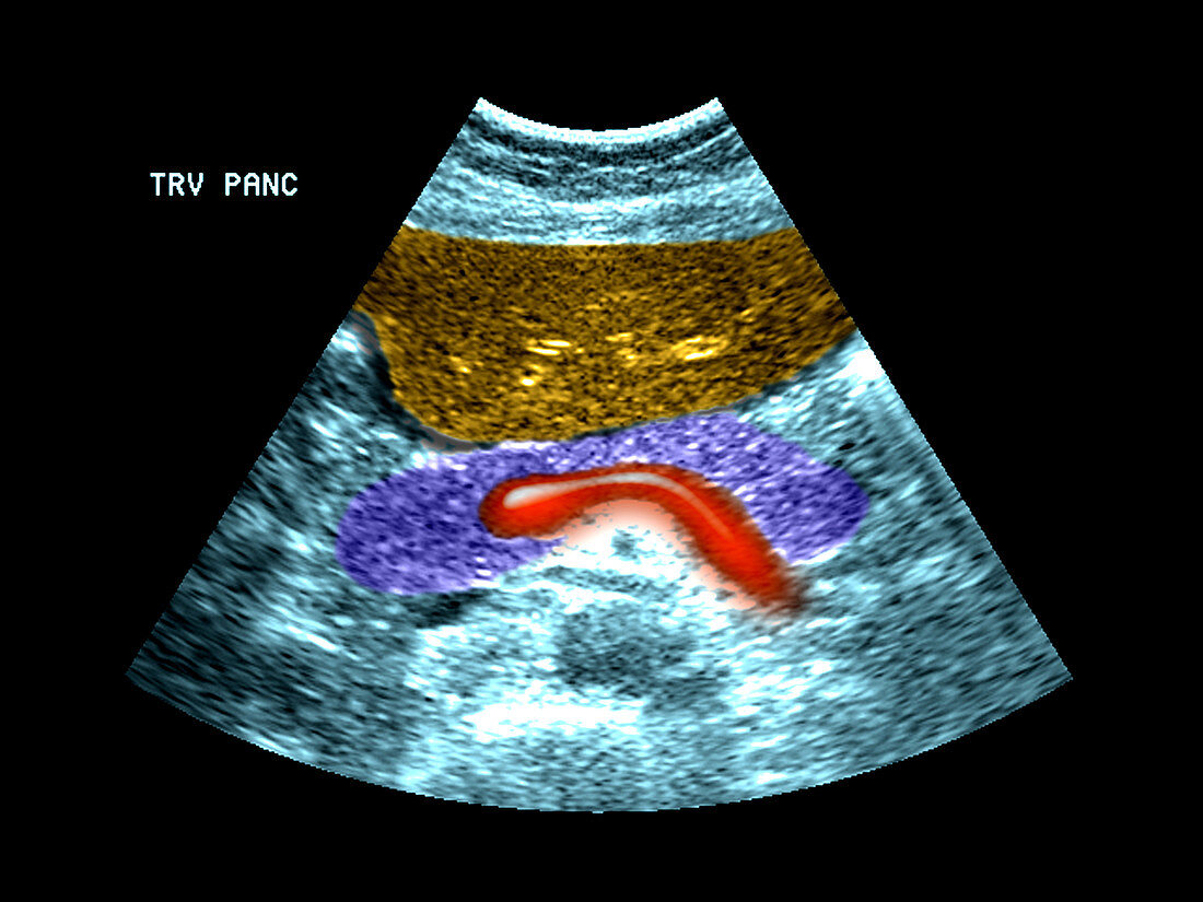
| This colour enhanced axial (cross section) ultrasound image of the epigastric region shows a nice example of the normal appearance of the pancreas (purple) anterior (towards the top) to the splenic vein (red). Near the top of the image is the left lobe of the liver (yellow) | |
| Licence : | Droits gérés |
| Crédit: | Science Photo Library / Living Art Enterprises, LLC |
| Taille de l’image : | 3600 px × 2700 px |
| Model Release : | Non requis |
| Property Release : | Non requis |
| Restrictions : |
|
Prix pour cette image À partir de 45 €
Produit vendu
(Calendrier, Carte postale, Carte de vœux, Impression sur textile, Packaging etc)
À partir de 45 €
Usage commercial
(Affichage, Annonce presse, Annonce TV, Carte, Digital - hors rés. sociaux, Digital - rés. sociaux etc)
À partir de 45 €
Éditorial
(Digital, Journal, Livre, Livre pratique, Magazine, Télévision etc)
À partir de 60 €
Usage non-commercial
(Digital - hors rés. sociaux, Digital - rés. sociaux etc)
À partir de 120 €
Mots clés
- anatomie,
- antérieur,
- axial,
- corps humain,
- couleur,
- couleur améliorée,
- couleur augmentée,
- coupe transversale,
- digestion,
- en bonne santé,
- épigastrique,
- foie,
- gut,
- image médicale,
- imagerie médicale,
- lobe,
- médical,
- médicale,
- normal,
- pancréas,
- précédent,
- sain,
- science,
- splénique,
- système digestif,
- ultrason,
- veine,
- veine splénique