CT of neck showing normal vocal chords
Numéro d’image : 12070788
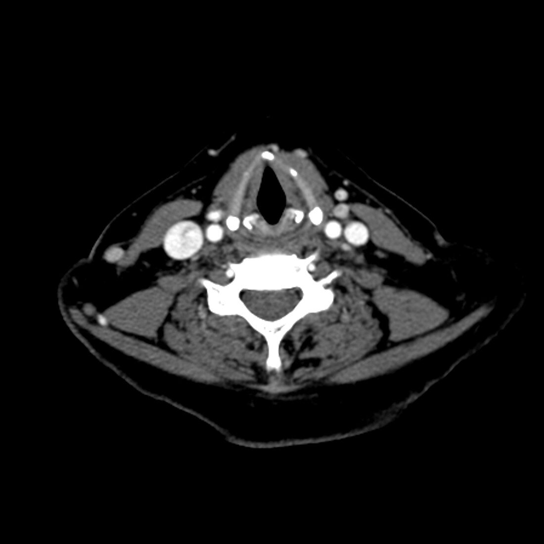
| This axial cross sectional CT image of the neck shows the appearance of normal vocal cords. The small white structures behind each vocal cord is the arytenoid cartilage. The carotid arteries and internal jugular veins are on either side of the neck. The spine is seen as the white structure in the middle of the image below the trachea and vocal cords | |
| Licence : | Droits gérés |
| Crédit: | Science Photo Library / Living Art Enterprises, LLC |
| Taille de l’image : | 6000 px × 6000 px |
| Model Release : | Non requis |
| Property Release : | Non requis |
| Restrictions : |
|
Prix pour cette image À partir de 45 €
Produit vendu
(Calendrier, Carte postale, Carte de vœux, Impression sur textile, Packaging etc)
À partir de 45 €
Usage commercial
(Affichage, Annonce presse, Annonce TV, Carte, Digital - hors rés. sociaux, Digital - rés. sociaux etc)
À partir de 45 €
Éditorial
(Digital, Journal, Livre, Livre pratique, Magazine, Télévision etc)
À partir de 60 €
Usage non-commercial
(Digital - hors rés. sociaux, Digital - rés. sociaux etc)
À partir de 120 €