Coronal MRI of Brachial Plexus
Numéro d’image : 12070573
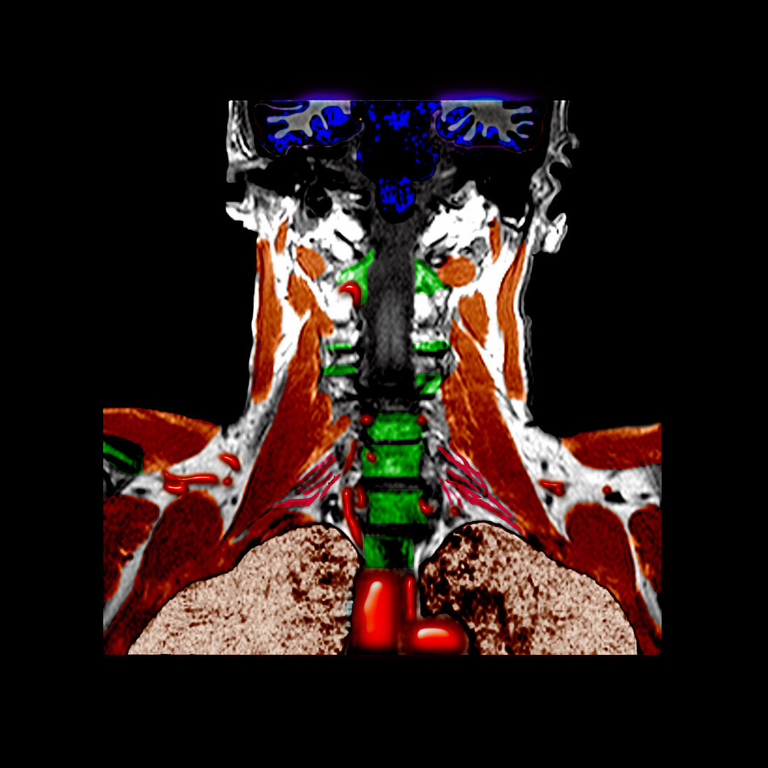
| colour enhanced,coronal MRI view done to evaluate the brachial plexus. The different neural components of the brachial plexus are the linear,stringlike red structures (towards the lower part of the image). Components of spinal nerve roots C4 to T1 contribute to the formation of the brachial plexus. The plexus extends out through the region of the shoulders and down both arms. The different components of the brachial plexus can be difficult to visualize due to their small size. The brown coloured structures represent different muscle groups. The neon green represents the cervical vertebrae. The bright red-orange structures in the lower part of the image are vascular structures. The largest ones are the superior vena cava on the left and the top of the aortic arch on the right | |
| Licence : | Droits gérés |
| Crédit: | Science Photo Library / Living Art Enterprises, LLC |
| Taille de l’image : | 3600 px × 3600 px |
| Model Release : | Non requis |
| Property Release : | Non requis |
| Restrictions : |
|
Prix pour cette image À partir de 45 €
Produit vendu
(Calendrier, Carte postale, Carte de vœux, Impression sur textile, Packaging etc)
À partir de 45 €
Usage commercial
(Affichage, Annonce presse, Annonce TV, Carte, Digital - hors rés. sociaux, Digital - rés. sociaux etc)
À partir de 45 €
Éditorial
(Digital, Journal, Livre, Livre pratique, Magazine, Télévision etc)
À partir de 60 €
Usage non-commercial
(Digital - hors rés. sociaux, Digital - rés. sociaux etc)
À partir de 120 €
Mots clés
- aiguillons,
- anatomie,
- central,
- colonne vertébrale,
- corps humain,
- cou,
- cous,
- diagnostic,
- diagnostique,
- dos,
- épines,
- I.R.M.,
- imagerie médicale,
- imagerie par résonance magnétique,
- imagerie par résonnance magnétique,
- IRM,
- moelle épinière,
- muscle,
- plexus brachial,
- S.N.C.,
- SNC,
- système nerveux,
- système nerveux central