Coloured CT scan of section through healthy brain
Numéro d’image : 12070519
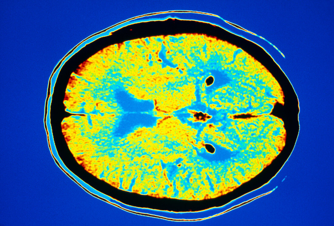
| Healthy brain. Coloured computed tomography (CT or CAT) scan of an axial section through a healthy brain from a 70-year-old man. The front of the brain is at left. The cerebral hemispheres are coloured yellow,with the ventricles blue. The black circles at upper and lower right on the ventricles either side of the brain's midline represent the choroid plexi of blood vessels. The pineal gland appears black and irregularly shaped between them. CT scanning combines the use of a computer and fine X-ray beams to produce images of "slices" through body parts. The scans produce good contrast between soft tissues and bone and are useful in diagnosing tumours and blood clots | |
| Licence : | Droits gérés |
| Crédit: | Science Photo Library / Camazine, Scott |
| Taille de l’image : | 3780 px × 2560 px |
| Model Release : | Non requis |
| Property Release : | Non requis |
| Restrictions : |
|
Prix pour cette image À partir de 45 €
Produit vendu
(Calendrier, Carte postale, Carte de vœux, Impression sur textile, Packaging etc)
À partir de 45 €
Usage commercial
(Affichage, Annonce presse, Annonce TV, Carte, Digital - hors rés. sociaux, Digital - rés. sociaux etc)
À partir de 45 €
Éditorial
(Digital, Journal, Livre, Livre pratique, Magazine, Télévision etc)
À partir de 60 €
Usage non-commercial
(Digital - hors rés. sociaux, Digital - rés. sociaux etc)
À partir de 120 €