Coloured TEM of skeletal (striated) muscle of fish
Numéro d’image : 12070113
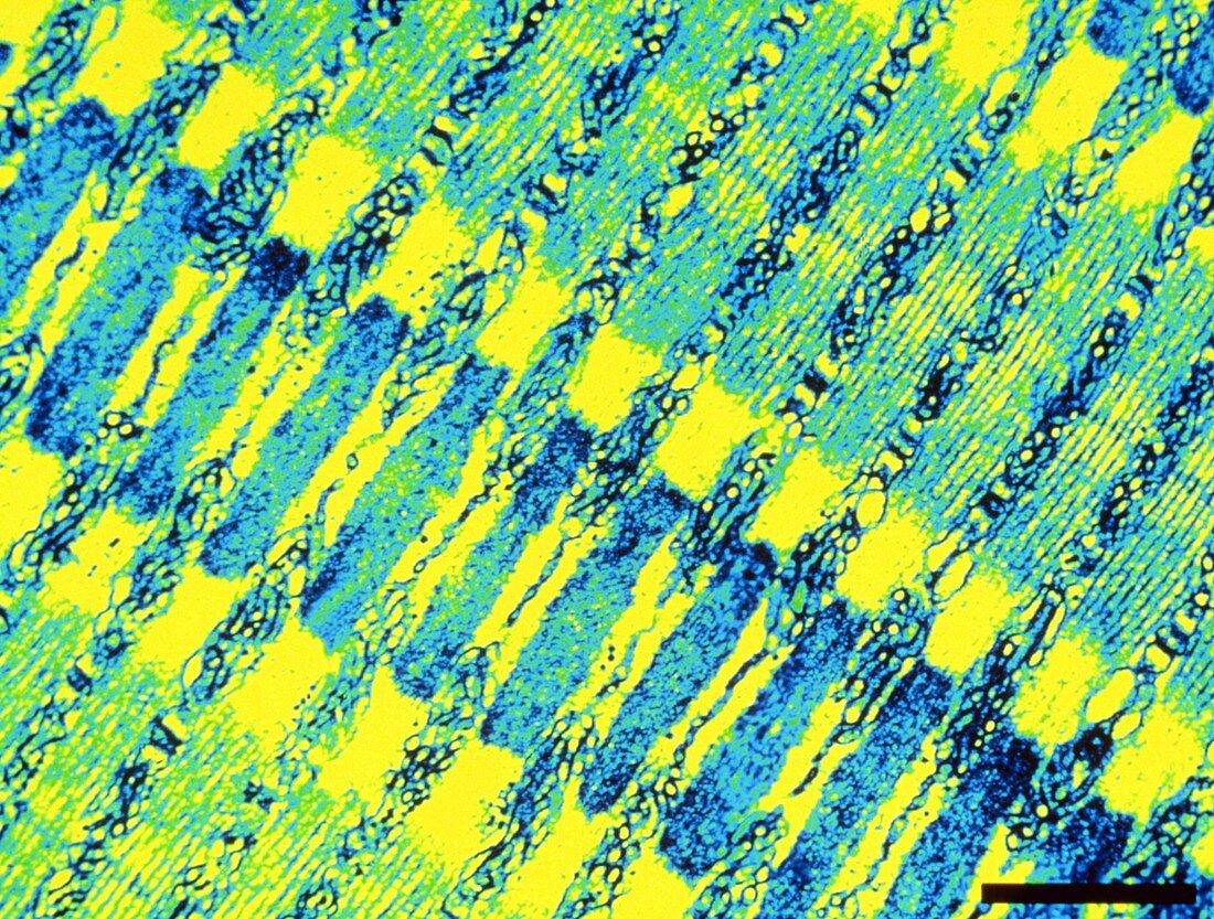
| Skeletal muscle of a fish. Coloured Transmission Electron Micrograph (TEM) of skeletal (striated) muscle of the fish Porichthys notatus. The banding pattern here gives striated muscle its name. These bands are formed by myofilaments (actin and myosin protein filaments). A wide Z-band (dark green) runs diagonally from upper left to bottom right,made of actin filaments. To its right is an A-band (light green) of thicker myosin filaments. These filaments slide over each other to cause muscle contraction. Sarcoplasmic reticulum (top right to bottom left) carries nerve impulses to these filaments. Magnification: x6,250 at 35mm size | |
| Licence : | Droits gérés |
| Crédit: | Science Photo Library / SCOTT CAMAZINE AND M. MARCHATERRE |
| Taille de l’image : | 4783 px × 3620 px |
| Model Release : | Non requis |
| Property Release : | Non requis |
| Restrictions : |
|
Prix pour cette image À partir de 45 €
Produit vendu
(Calendrier, Carte postale, Carte de vœux, Impression sur textile, Packaging etc)
À partir de 45 €
Usage commercial
(Affichage, Annonce presse, Annonce TV, Carte, Digital - hors rés. sociaux, Digital - rés. sociaux etc)
À partir de 45 €
Éditorial
(Digital, Journal, Livre, Livre pratique, Magazine, Télévision etc)
À partir de 60 €
Usage non-commercial
(Digital - hors rés. sociaux, Digital - rés. sociaux etc)
À partir de 120 €