Cardiac muscle with intercalated discs
Numéro d’image : 12070112
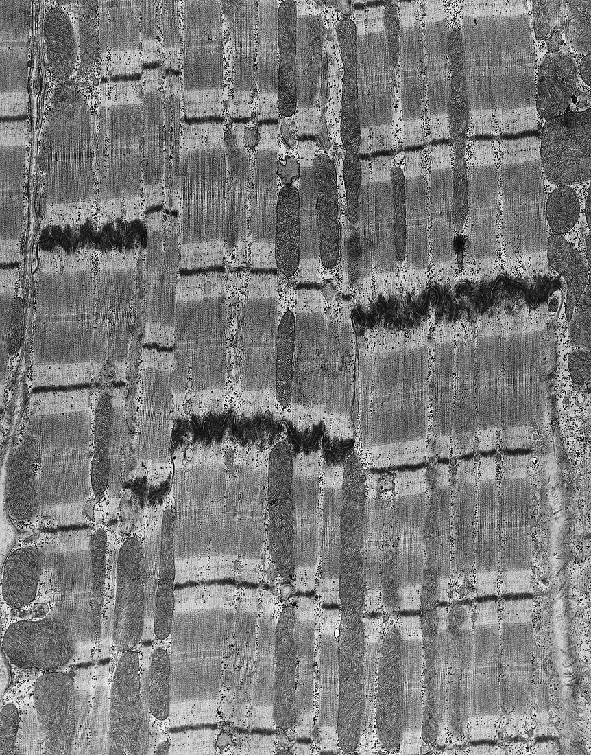
| Transmission electron micrograph of cardiac muscle showing 3 intercalated discs (thick dark stained bands). These are specialised junctions between cardiac muscle cells,providing areas of low electrical resistance for the rapid spread of excitation through the muscle. The elongated bodies between the muscle fibres are mitochondria,energy-rich sites which fuel muscle activity. The black dots are glycogen,sites of carbohydrate storage. A cell contains two types of myofibrils; dark bands of myosin filaments & light bands of actin filaments. These filaments are thought to slide over each other,fuelled by energy released from ATP,causing the muscle to contract | |
| Licence : | Droits gérés |
| Crédit: | Science Photo Library / Fawcett, Dr. Don |
| Taille de l’image : | 3952 px × 5041 px |
| Model Release : | Non requis |
| Property Release : | Non requis |
| Restrictions : |
|
Prix pour cette image À partir de 45 €
Produit vendu
(Calendrier, Carte postale, Carte de vœux, Impression sur textile, Packaging etc)
À partir de 45 €
Usage commercial
(Affichage, Annonce presse, Annonce TV, Carte, Digital - hors rés. sociaux, Digital - rés. sociaux etc)
À partir de 45 €
Éditorial
(Digital, Journal, Livre, Livre pratique, Magazine, Télévision etc)
À partir de 60 €
Usage non-commercial
(Digital - hors rés. sociaux, Digital - rés. sociaux etc)
À partir de 120 €