LM of normal human cardiac muscle fibres
Numéro d’image : 12070087
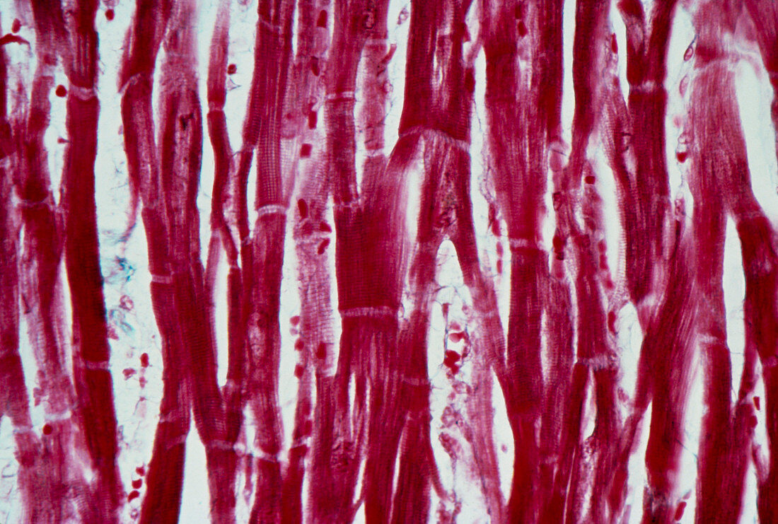
| Light micrograph of human cardiac muscle fibres,shown in longitudinal section (LS). The cells contain 1-2 nuclei and an extensive branching cytoplasm (synctium). The nuclei are centrally located (in skeletal muscle they are located below the surface of the sarcolemma,the membrane surrounding the muscle fibres),and the regular cross-striations are visible. The intracellular spaces are filled with connective tissue,rich in blood vessels: this reflects the demands on cardiac muscle of strong & continuous contractility. Mag. X 106 (35mm) | |
| Licence : | Droits gérés |
| Crédit: | Science Photo Library / Abbey, Michael |
| Taille de l’image : | 3682 px × 2480 px |
| Model Release : | Non requis |
| Property Release : | Non requis |
| Restrictions : |
|
Prix pour cette image À partir de 45 €
Produit vendu
(Calendrier, Carte postale, Carte de vœux, Impression sur textile, Packaging etc)
À partir de 45 €
Usage commercial
(Affichage, Annonce presse, Annonce TV, Carte, Digital - hors rés. sociaux, Digital - rés. sociaux etc)
À partir de 45 €
Éditorial
(Digital, Journal, Livre, Livre pratique, Magazine, Télévision etc)
À partir de 60 €
Usage non-commercial
(Digital - hors rés. sociaux, Digital - rés. sociaux etc)
À partir de 120 €