Compact bone
Numéro d’image : 12069904
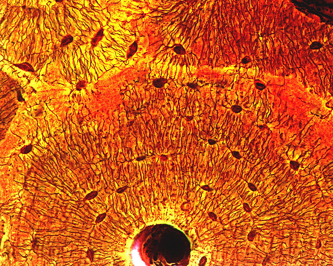
| Light micrograph showing a section through compact bone of a mature long bone at high magnification. The micrograph shows in cross section half of a Haversian system and includes its Haversian canal. The osteocytes appear as densely stained,elongate cell bodies from which numerous cytoplasmic processes extend to meet the cytoplasmic processes of cells in adjacent lamellae. The upper portion of the micrograph reveals portions of interstitial lamellae. Schmorls stain. Magnification: x700 | |
| Licence : | Droits gérés |
| Crédit: | Science Photo Library / Ross, Michael |
| Taille de l’image : | 1880 px × 1500 px |
| Model Release : | Non requis |
| Property Release : | Non requis |
| Restrictions : |
|
Prix pour cette image À partir de 45 €
Produit vendu
(Calendrier, Carte postale, Carte de vœux, Impression sur textile, Packaging etc)
À partir de 45 €
Usage commercial
(Affichage, Annonce presse, Annonce TV, Carte, Digital - hors rés. sociaux, Digital - rés. sociaux etc)
À partir de 45 €
Éditorial
(Digital, Journal, Livre, Livre pratique, Magazine, Télévision etc)
À partir de 60 €
Usage non-commercial
(Digital - hors rés. sociaux, Digital - rés. sociaux etc)
À partir de 120 €