Woman undergoing PET scan
Numéro d’image : 12068618
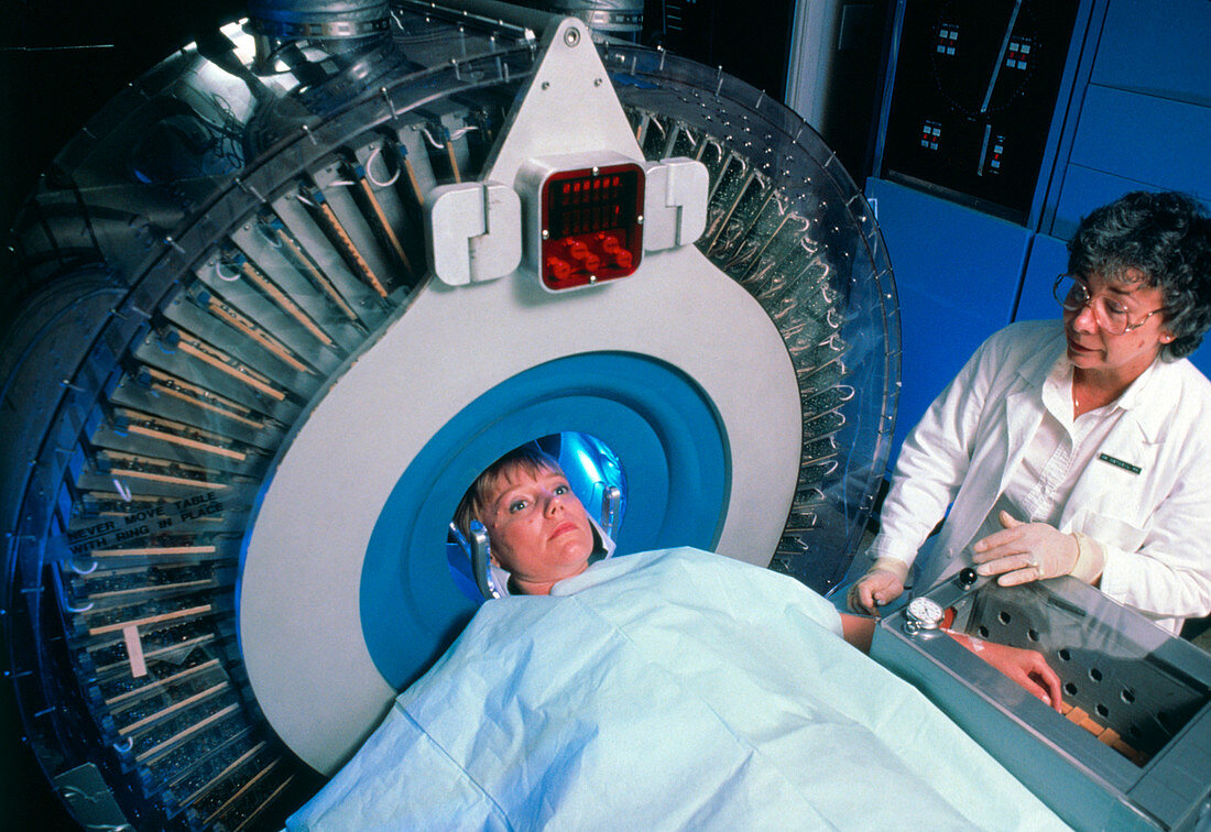
| MODEL RELEASED. Woman undergoing brain scan using Positron Emission Tomography (PET),showing her head surrounded by the circular detector. A colour- coded PET scan is obtained by first injecting a positron-emitting radioisotope (produced in a cyclotron) into the bloodstream: the detector is sensitive to gamma rays produced when positrons emitted annihilate with electrons from the molecules in the brain (known as tracers) to which the radioisotope has been dedicated. Applications of PET include the use of radioactive oxygen to measure oxygen consumption in senile dementia,cerebrovascular disease and brain tumours | |
| Licence : | Droits gérés |
| Crédit: | Science Photo Library / Morgan, Hank |
| Taille de l’image : | 3661 px × 2517 px |
| Model Release : | Disponible |
| Property Release : | Non requis |
| Restrictions : |
|
Prix pour cette image À partir de 45 €
Produit vendu
(Calendrier, Carte postale, Carte de vœux, Impression sur textile, Packaging etc)
À partir de 45 €
Usage commercial
(Affichage, Annonce presse, Annonce TV, Carte, Digital - hors rés. sociaux, Digital - rés. sociaux etc)
À partir de 45 €
Éditorial
(Digital, Journal, Livre, Livre pratique, Magazine, Télévision etc)
À partir de 60 €
Usage non-commercial
(Digital - hors rés. sociaux, Digital - rés. sociaux etc)
À partir de 120 €